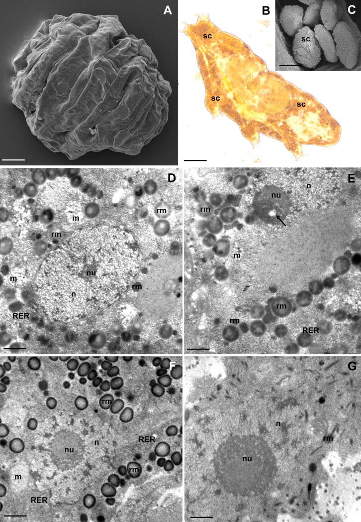
Fig 1. Storage cells (SC) of R. coronifer.
(A) Tun, SEM. Bar = 30 μm. (B) Active animal, LM. Bar = 20 μm. (C) Storage cells, SEM. Bar = 4 μm. (D-G) Ultrastructure of SC of non-experimental specimens, TEM: nucleus (n), nucleolus (nu), mitochondria (m), rough endoplasmic reticulum (RER), spheres of reserve material (rm). (D-E). SC of male specimens. (D) Bar = 0.58 μm. (E) Bar = 0.5 μm. (F-G) SC of female specimens. (F) SC of the first type during vitellogenesis, nucleolus vacuole (arrow). Bar = 0.8 μm. (G) SC of the second type. Bar = 0.5 μm.

