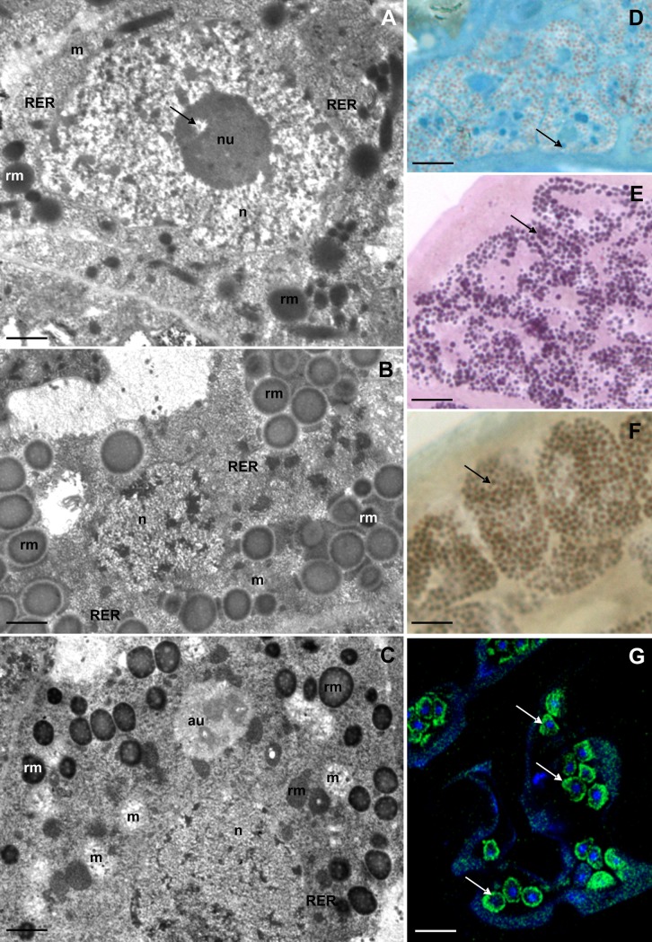
Fig 2. Ultrastructure and histochemistry of the SC of the first type during different stages of oogenesis.
(A-C) Ultrastructure of SC, TEM: nucleus (n), nucleolus (nu), mitochondria (m), rough endoplasmic reticulum (RER), spheres of reserve material (rm). (A) Previtellogenesis, nucleolus vacuole (arrow). Bar = 0.47 μm. (B) Late vitellogenesis. Bar = 0.57 μm. (C) Late choriogenesis, autophagosome (au). Bar = 0.65 μm. (D-G). Histochemical staining of SC, arrow indicates positive reaction: (D) BPB staining, LM. Bar = 4 μm. (E) PAS method, LM. Bar = 3.5 μm. (F) Sudan Black B staining, LM. Bar = 3 μm. (G) BODIPY 493/503 and DAPI staining, confocal microscopy. Bar = 10 μm.

