
Figure 1. Histological assessment of DRG tissue.
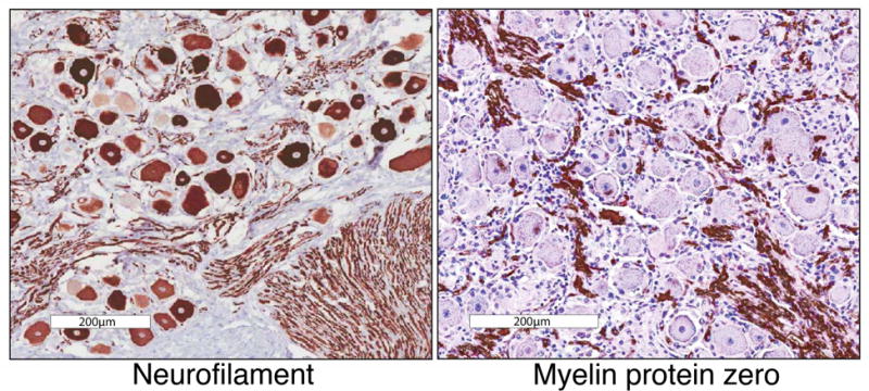
DRG tissue was stained for either neurofilamen light chain (left) or myelin protein zero (right), which are markers for neurons and Schwann cells respectively. In the case of neurofilament, these proteins are made in the soma, but functional assembly is also required for axonal structure. Because of this, this protein stains both neuronal cell bodies and axonal bundles. These genes are among the most highly expressed, highly differential genes in each tissue. The presence of myelin protein tracks in the DRG is indicative of Schwann cell coated myelinated axonal sheaths extending out from the DRG into the dorsal roots, and to the periphery and the spinal cord. These cells represent a major non-neuronal component of DRGs. Scale bar represents 200μm.