
Figure 2.
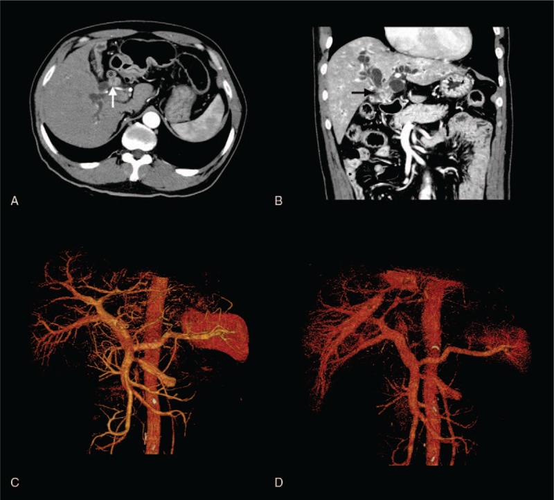
Abdominal enhanced computed tomography (CT) images. (A, B) An enhanced CT scan showed a moderately enhanced mass located in the hilar bile duct (white and black arrows) with regional lymph node involvement (asterisk). (C, D) CT angiography and 3-dimensional reconstruction revealed the absence of vascular invasion.