
Figure 3.
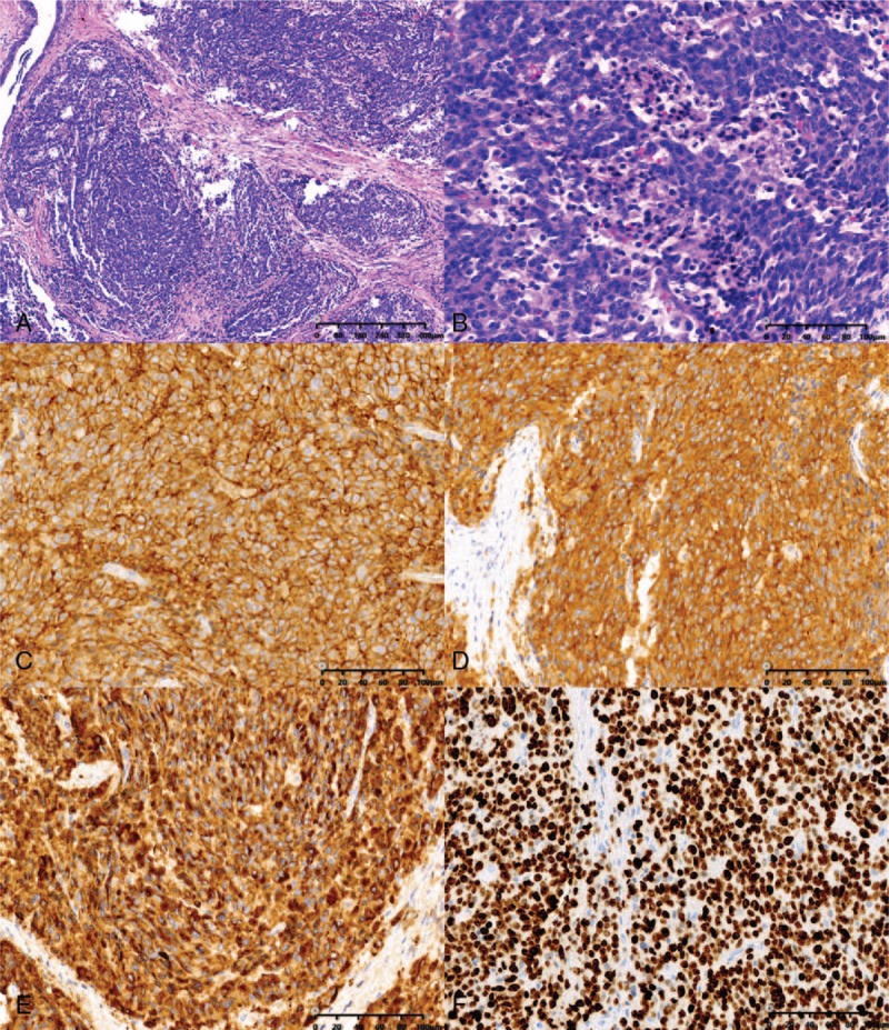
Microscopy images of the surgical specimen. (A) The tumor showed a nested organoid growth pattern (hematoxylin and eosin [HE], ×100). (B) The tumor cells were small in size and had round, hyperchromatic nuclei and scant cytoplasm (HE, ×400); immunohistochemical examinations revealed that the tumor cells were positive for CD56 (C, ×400), synaptophysin (D, ×400), and chromogranin (E, ×400); more than 80% of the tumor cells were positive for Ki-67 (F, ×400).