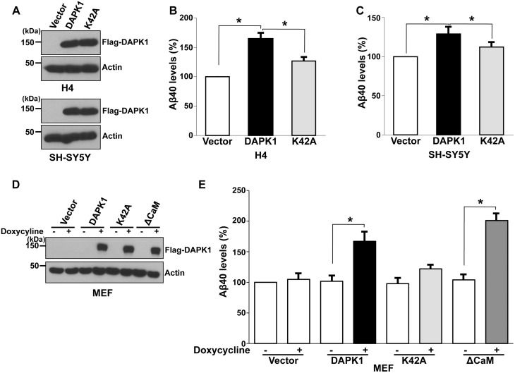
Figure 1.
DAPK1 stimulates the secretion of human- and mouse Aβ40. (A–C) Human H4 and SH-SY5Y cells were transfected with pRK5-Flag, pRK5-Flag-DAPK1 or pRK5-Flag-DAPK1K42A for 32 h. The cell lysates were subjected to western blot analysis with anti-Flag or anti-actin antibody and the levels of human Aβ40 in cell culture supernatants were determined by a solid phase sandwich ELISA assay. Each data point represents the mean ± standard error of three independent experiments (*P < 0.05; ANOVA/Dunnett’s test). (D, E) MEF cells were transfected with pTRE-Tight vector, pTRE-Tight-DAPK1, pTRE-Tight-DAPK1K42A or pTRE-Tight-DAPK1ΔCaM for 8 h followed by treatment with or without 1 μg/ml doxycycline for 48 h. The cell lysates were subjected to western blot analysis with anti-Flag or anti-actin antibody and the levels of mouse Aβ40 in cell culture supernatants were determined by a solid phase sandwich ELISA assay. Each data point represents the mean ± standard error of three independent experiments (*P < 0.05; ANOVA/Dunnett’s test).

