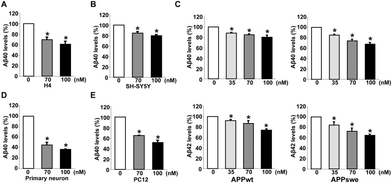
Figure 3.
The pharmacological inhibitor of DAPK1 reduces the secretion of Aβ40 and Aβ42. (A, B) Human H4 (A) and SH-SY5Y (B) cells were treated with the indicated concentrations of DAPK1 pharmacological inhibitor for 24 h. Levels of human Aβ40 in cell culture supernatants after 24 h incubation were determined by a solid phase sandwich ELISA assay as in (B) and (C). Each data point represents mean ± standard error of three independent experiments (*P < 0.05; ANOVA/Dunnett’s test). (C) SH-SY5Y APPwt and SH-SY5Y APPswe cells were incubated with the indicated concentrations of DAPK1 pharmacological inhibitor for 24 h. The supernatants were subjected to human Aβ40 and Aβ42 ELISA analyses as in (C). Each data point represents mean ± standard error of three independent experiments (*P < 0.05; ANOVA/Dunnett’s test). (D, E) Primary cortical neurons (D) at DIV 8–10 and the rat adrenal pheochromocytoma PC12 (E) cells were treated with the indicated concentrations of DAPK1 pharmacological inhibitor for 36 h. Levels of Aβ40 in cell culture supernatants after 36 h incubation were determined by a solid phase sandwich ELISA assay. Each data point represents mean ± standard error of three independent experiments (*P < 0.05; ANOVA/Dunnett’s test).

