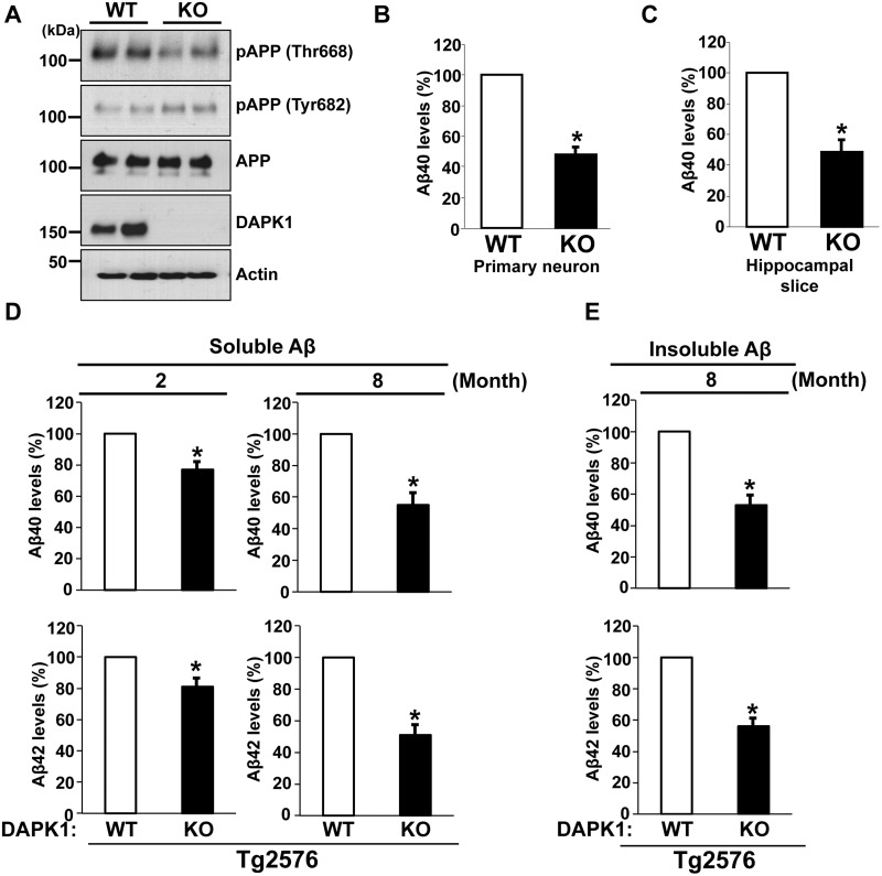
Figure 6.
DAPK1 KO reduces the secretion of Aβ40 and Aβ42 in mouse models. (A) Whole brain lysates from WT and DAPK1 KO mice harvested at 6 month of age were analyzed for the levels of phosphorylated (Thr668 and Tyr682) and total APP protein as well as DAPK1. Anti-actin antibody was used as a loading control. The blots are representative of three independent experiments. Analyzed mice number: WT (n = 6, 3 male (M) and 3 female (F)), DAPK1 KO (3M, 3F). (B, C) Primary cortical neurons (B) at DIV 8-10 of WT and DAPK1 KO mice and the brain hippocampal tissue slices (C) prepared from WT and DAPK1 KO mice at 6 month of age were cultured for 36 h. Levels of mouse Aβ40 in cell culture supernatants after 36 h incubation were determined by a solid phase sandwich ELISA assay. Each data point represents mean ± standard error of three independent experiments (*P < 0.05; ANOVA/Dunnett’s test). (D) TBS-buffer soluble hippocampal tissue fraction from Tg2576/WT or Tg2576/DAPK1 KO mice at 2 or 8 month of age was prepared. Levels of human Aβ40 and Aβ42 were determined by a solid phase sandwich ELISA assay. Each data point represents mean ± standard error of three independent experiments (*P < 0.05; ANOVA/Dunnett’s test). Analyzed mice number, 2-month-old groups: Tg2576/WT (3M, 3F), Tg2576/DAPK1 KO (3M, 3F); 8-month-old groups: Tg2576/WT (3M, 3F), Tg2576/DAPK1 KO (3M, 3F). (E) TBS-buffer insoluble hippocampal tissue fraction from Tg2576/WT or Tg2576/DAPK1 KO mice at 8 month of age was prepared. Levels of human Aβ40 and Aβ42 were determined by a solid phase sandwich ELISA assay. Each data point represents mean ± standard error of three independent experiments (*P < 0.05; ANOVA/Dunnett’s test). Analyzed mice number: Tg2576/WT (3M, 3F), Tg2576/DAPK1 KO (3M, 3F).

