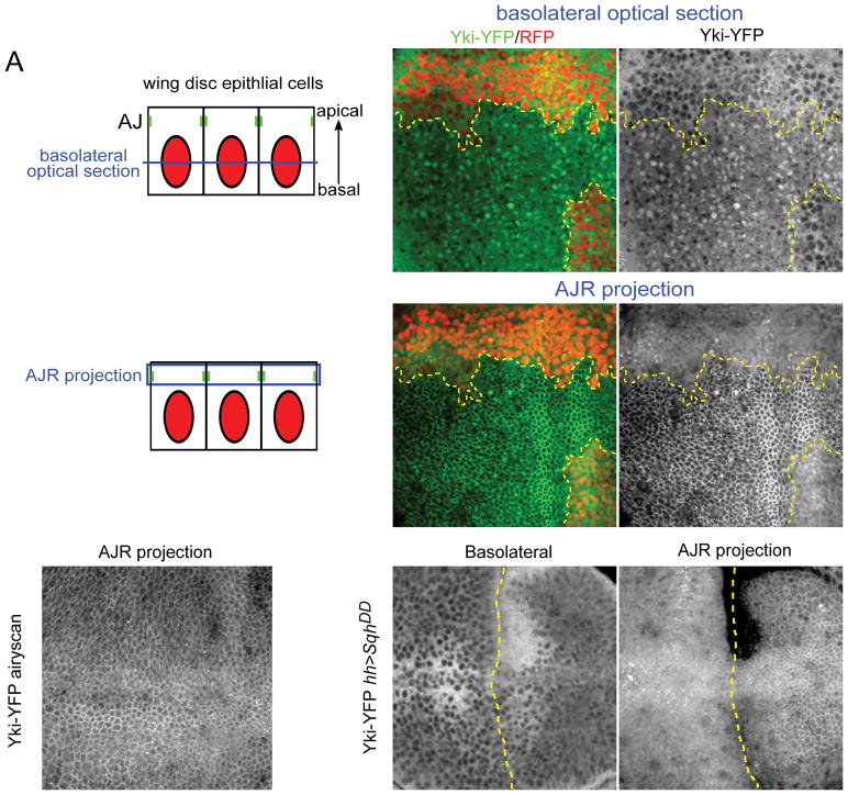
Figure 1. Yki localizes to the apical junctional region (AJR) in addition to the nucleus.
(A) Illustrations showing cross-sectional views of the imaginal epithelium and the approximate positions of basolateral and AJR images shown in this study. As indicated in blue, basolateral images are single sections while AJR views are maximal projections of a small number of apical sections to compensate for curvature of the epithelium.
(B–C′) The effect of Hippo pathway inactivation on Yki subcellular localization. In basal sections of live tissues containing wts null mitotic clones (clone marked by the absence of RFP and a yellow dashed line), Yki-YFP is primarily cytoplasmic in normal imaginal tissue, but is strongly nuclear in wts mutant cells (B–B′). Apically, Yki-YFP is slightly enriched at the apical cortex in normal tissues, but strongly recruited to the AJR in wts null clones (C–C′).
(D) Yki localizes to the AJR in normal tissues. A super-resolution image (using the Zeiss Airyscan that improves resolution and signal-to-noise) of a live wing disc carrying two copies of endogenously expressed Yki-YFP in a ykiB5 null background. A maximum projection of apical optical sections shows cortical localization of Yki-YFP apically.
(E–F) The effect of increasing cytoskeletal tension on Yki subcellular localization. Decreasing Hippo pathway activity by increasing cytoskeletal tension in cells that express a constitutively active form of the myosin regulatory light chain, SqhDD, caused increased nuclear and AJR localization of Yki-YFP. The boundary of SqhDD expression, under the control of hh>Gal4, is marked with a dashed yellow line.

