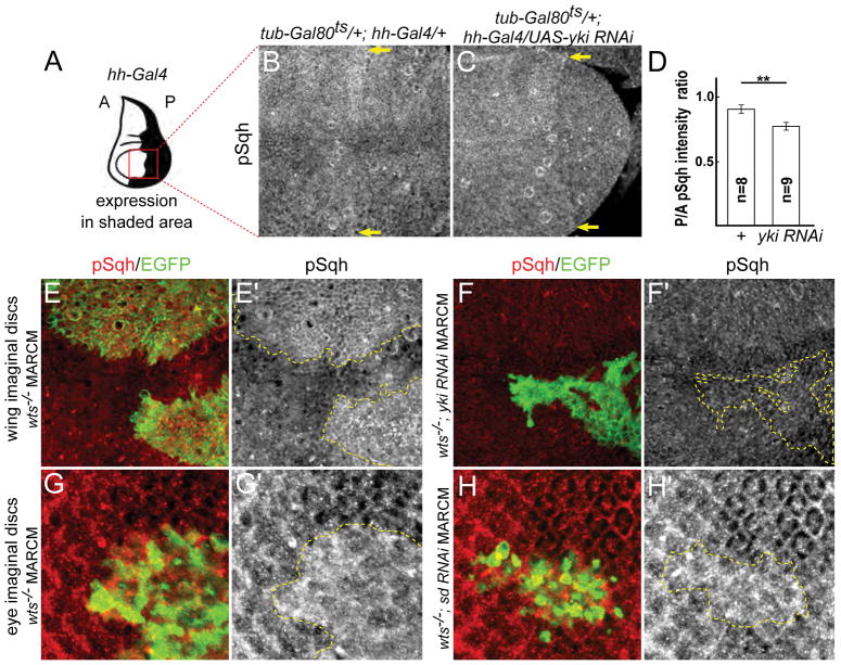
Figure 3. Yki promotes myosin activation in normal tissues and in response to Hippo pathway inactivation.
(A) Cartoon showing expression domain of hh-Gal4 in the wing disc. Red box indicates the approximate area of the wing shown in B–C. A: anterior, P: posterior.
(B–C) Depletion of Yki results in decreased Sqh activation. Expression of a yki-RNAi transgene for two days prior to dissection results in decreased pSqh staining, indicating Yki normally promotes Sqh activation. Images are maximal projections of apical optical sections. Yellow arrows indicate the hh-Gal4 expression boundary.
(D) Quantification of the ratio of pSqh staining fluorescence intensity between posterior (P) and anterior (A) compartments. There is a significant reduction of pSqh ratio in yki RNAi wing discs. Data are represented as mean ± SEM. Asterisks represent statistical significance of the difference between selected groups (** p<0.01, One-way ANOVA and Tukey’s HSD test, n = number of wing discs).
(E–F′) Yki promotes myosin activation in response to Hippo pathway inactivation. pSqh staining was increased in wts null (wtsX1) mitotic clones in wing discs (E–E′; clone marked with GFP and yellow dashed lines), suggesting Yki promotes myosin activation upon Hippo pathway inactivation. This increased pSqh staining is suppressed when yki is depleted in wts null clones (F–F′; clone marked with GFP and yellow dashed lines), suggesting endogenous Yki can promote myosin activation.
(G–H′) Yki promotes myosin activation independent of its transcriptional function. pSqh staining was increased in wtsX1 null mitotic clones in eye discs (G–G′; clone marked with GFP and yellow dashed lines). This increased pSqh staining is maintained when sd is depleted in wts null clones (H–H′; clone marked with GFP and yellow dashed lines), suggesting Yki promotes myosin activation independent of its transcriptional function.
See also Figure S2.

