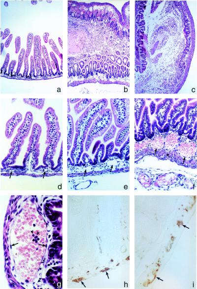
Figure 3.
Jejuno-ileo-colitis develops in (GFAP-HA × CL4-TCR)F1 double transgenic mice. (a) Hematoxylin/eosin-stained section showing normal jejunal histology in a 7-day-old GFAP-HA transgenic mouse. Late-stage lesion of the jejunum (b) and colon (c) in double transgenic mice with mucosal and submucosal edema, epithelial damage, mucosal inflammation, hemorrhage, and necrosis that severely perturbed the crypt-villous architecture and mucosal integrity (×40 for a–c). (d) Normal jejunal histology in GFAP-HA trangenic mice. (e and f) Pathology in double transgenic mice was initially observed in submucosal capillaries and blood vessels (arrows) (×100 for d–f); asterisk in f indicates smooth muscle thickening; (g) with vasodilation, erythrostasis, endothelial hypertrophy and hyperplasia (arrows), and neutrophil (arrowheads) and lymphocyte infiltration. (h) PGP9.5 immunostaining in the jejunal myenteric plexus in GFAP-HA transgenic and (i) double transgenic mice (×400 for g–i).

