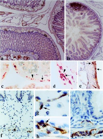
Figure 4.
Small intestine pathology in (GFAP-HA × CL4-TCR)F1 double transgenic mice. (a) Five-day-old double transgenic pup with moderate inflammation in the gut wall, associated with edema and vasodilatation. Inflammation (anti-CD3) was also observed in the surrounding mesentery. (b) Nontransgenic littermate; normal gut architecture; T cells are concentrated mainly in lymph follicles (×90 for a and b). (c and d) Double transgenic animal, infiltration of the myenteric plexus with T cells (c, ×360; d, ×720); double staining with anti-CD3 (brown, arrows) and GFAP (red, arrowheads). (e) Double transgenic animal; immunohistochemistry with anti-CD8; infiltration of the muscular wall and the myenteric plexus by CD8+ T cells (×360). (f) Double transgenic animal; GFAP+ immunohistochemistry; edema and inflammation in the mucosa and submucosa; only a few GFAP+ cells remain in the myenteric plexus (×450). (g) Double transgenic animal; apoptotic GFAP+ cell in the submucosal plexus (×1,300). (h) Double transgenic animal; apoptotic GFAP+ cell in the myenteric plexus (×1,300). (i) GFAP+ enteric glial cells in the myenteric and submucosal plexi in a normal, nontransgenic animal (×1,000).

