
Figure 3: Nucleation barriers govern prion behavior.
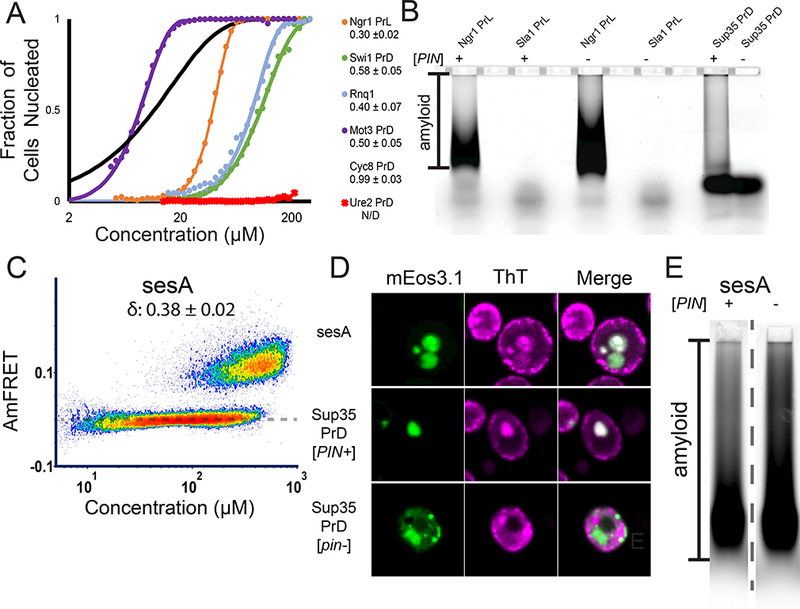
(A) Fits of DAmFRET of known amyloid-forming yeast polypeptides that exhibit bimodal distributions. The inset shows δ ± error. All fits are of [PIN+] cells, except for Rnq1. See also Fig. S3 A, B.
(B)SDD-AGE of Ngr1 PrL and Sla1 PrL in [PIN+] or [pin−] cells. Alternate lanes were intentionally left blank. See also Fig. S3C.
(C) DAmFRET of sesA, showing a bimodal distribution characteristic of prions.
(D) Representative images of ThT-stained cells expressing sesA, positive control Sup35 PrD in [PIN+] and negative control Sup35 PrD in [pin−].
(E) SDD-AGE of sesA in [PIN+] and [pin−] cells. The dashed line indicates the two adjacent lanesshown are spliced from different positions in the gel. See also Fig. S3D.