
Figure 4. A large nucleation barrier allows the archetypal prion protein, Sup35 PrD, to partition into physiological mRNP condensates.
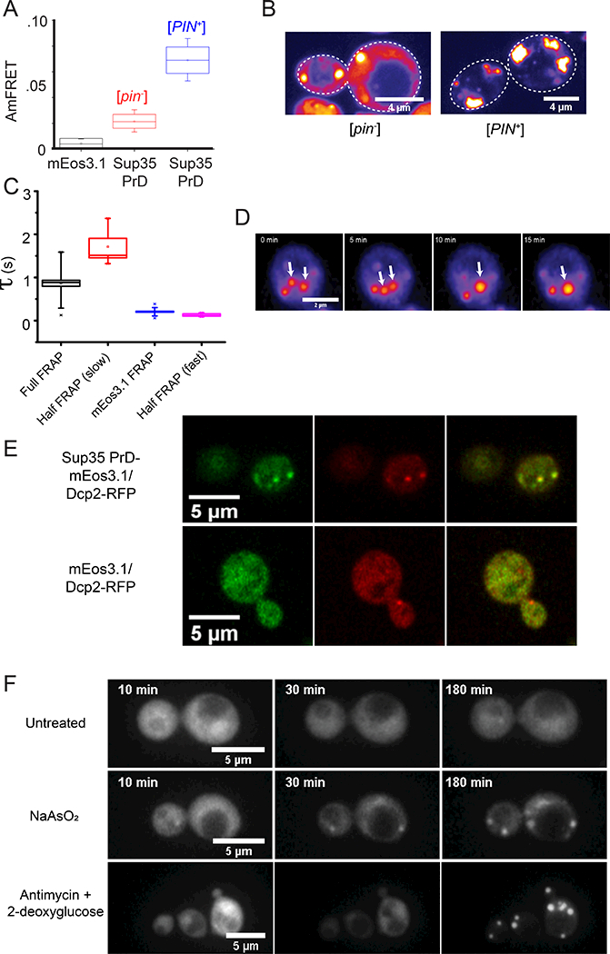
(A) Box-Whisker plot showing mean AmFRET of Sup35 PrD. The box is the SD of the mean, and the whiskers are the 5th and 95th percentiles of AmFRET values, for more than 2000 cells expressing between 80 and 200 μM of either unfused mEos3.1 (black) or Sup35 PrD in [pin−] (red) or [PIN+] cells (blue). See also Fig. S4A.
(B) Representative confocal images of Sup35 PrD puncta in [pin−] (left) or [PIN+] cells (right).
(C) Fluorescence recovery timescales of Sup35 PrD puncta in [pin−] cells as measured by FRAP. Error bars represent SD.
(D) Time-lapse microscopy showing coalescence of Sup35 PrD puncta. Single confocal slice of a [pin−] cell expressing Sup35 PrD, showing two puncta (white arrows from 0–5 min) coalesce into one larger punctum (single arrow from 10 min). See also Fig. S4B.
(E) Images of single confocal slices of cells co-expressing Sup35 PrD fused with mEos3.1 (and unfused mEos3.1 as control) and RFP-fused Dcp2 at 100X magnification.
(F) Montage of cells expressing Sup35 PrD showing the formation of liquid droplets upon treatment with NaAsO2 (middle row) or antimycin + 2-deoxyglucose (bottom), versus untreated cells (top). See also Movies S2–4.