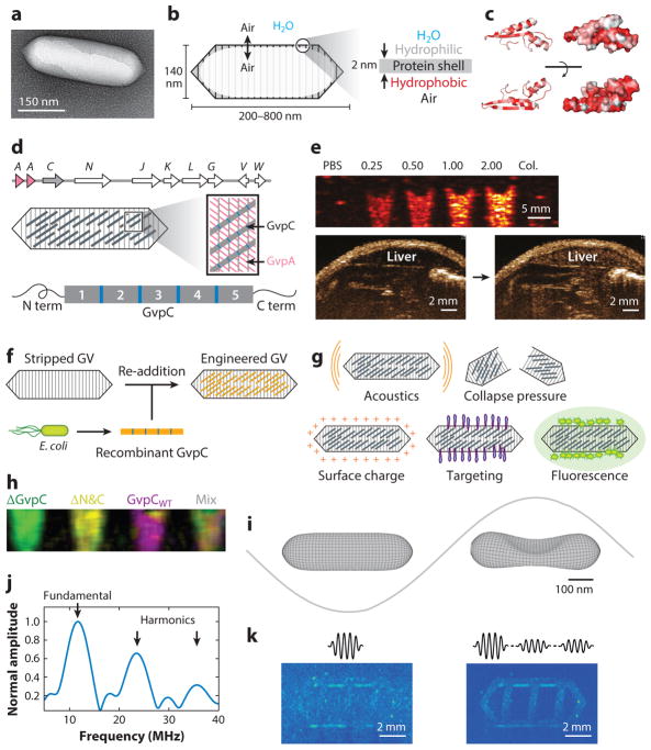
Figure 3.
Gas vesicles as acoustic biomolecules. (a) Transmission electron micrograph of individual gas vesicle (GV) from Anabaena flos-aquae. (b) Illustration of GV structure. (c) Protein folding model for A. flos-aquae gas vesicle protein A (GvpA), colored to indicate hydrophobilicy (red). Structure from Ezzeldin et al. (40), rendered in PyMOL. (d) Gene cluster encoding A. flos-aquae GVs (top), illustration of GvpA and GvpC spatial arrangement (middle), and repeat structure of GvpC protein (bottom). (e) Ultrasound image of GVs at various optical densities and after hydrostatic collapse in vitro. Image of mouse during and after GV administration in vivo, showing contrast in the liver owing to GV accumulation. (f) Illustration of protocol for replacing native GvpC with engineered versions. (g) GV properties that can be modified by substituting engineered versions of GvpC. (h) Multiplexed image of engineered GVs acquired with pressure spectral unmixing. (i) Finite element model of A. flos-aquae GV buckling under acoustic pressure. (j) Predicted acoustic output from buckling GVs. (k) B-mode and amplitude modulated images of gas vesicles arranged in a phantom with linear background scatterers. Panel a adapted with permission from Lu et al. (143). Panels d, f–h adapted with permission from Lakshmanan et al. (43). Panel e adapted with permission from Shapiro et al. (35). Panels i–k adapted with permission from Maresca et al. (45).

