
Fig. 1. Typical morphology and structure characterization results of Te nanoparticles prepared by ns-LAL.
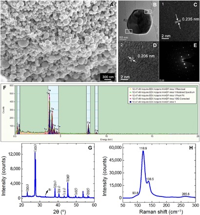
(A) SEM image of Te nanoparticles. (B) TEM image of a Te nanoparticle. (C and D) Corresponding HRTEM micrographs. (E and F) SAED and EDS patterns of the Te nanoparticle. (The Cu signal in EDS pattern originated from the Cu grid.) (G) XRD pattern of Te nanoparticles deposited on a Si substrate. (H) Raman spectrum of Te nanoparticles.