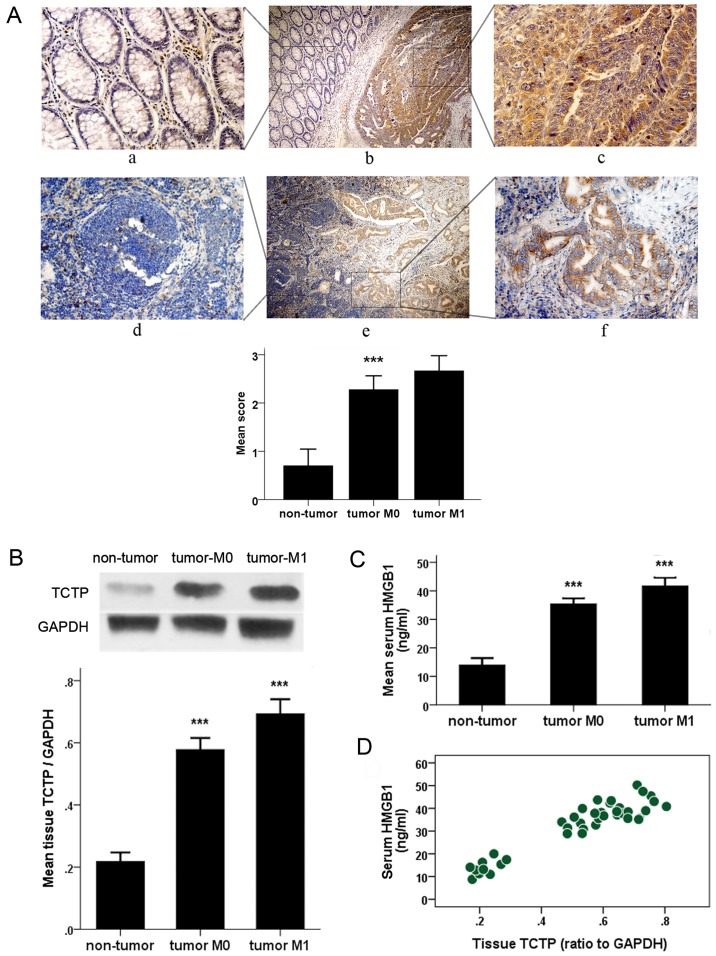
Figure 1.
Detection of the histological expression of TCTP and serum value of HMGB1 in patients with CRC. (A) Immunohistochemical detection of expression of TCTP in CRC tissues (upper panel) and metastatic lymph nodes (lower panel) and the adjacent normal tissues. Specific TCTP staining is shown in brown. Panels a, c, d and f, show a magnification of ×400; panels b and e show a magnification of ×100. Panels a, c, d and f are amplifications of the marked (with squares) areas shown in panels b and e, respectively (***P<0.001 between CRC and non-tumor groups). (B) Immunoblot analysis of expression of TCTP in colorectal samples from patients with CRC or from control subjects with benign polyps (non-tumor). Values were normalized to the internal control GAPDH (***P<0.001 between each two groups). (C) Enzyme-linked immunoassay examination of serum levels of HMGB1 in patients with CRC and control subjects (non-tumor) (***P<0.001 between each two groups). (D) Correlation plot generated with serum HMGB1 values and tumor tissue TCTP expression in 30 patients with CRC and 10 non-cancerous controls (Pearson's correlation coefficient, 0.935; P<0.001). CRC, colorectal cancer; TCTP, translationally controlled tumor protein; HMGB1, high mobility group box 1; M0, no metastasis; M1, metastasis.

