. 2018 Aug 10;7:e38461. doi: 10.7554/eLife.38461

Figure 1. Overall architecture of homomeric SWELL1.

(A) Cryo-EM reconstruction of SWELL1 homohexamer viewed from the membrane plane highlighting a dimer pair (top left, red and pink subunits) and an interface between dimers (top right, pink and green subunits), from the extracellular side (bottom left), and from the cytosolic side (bottom right). (B) Detailed view of SWELL1 ‘inner’ protomer. (C) Topology diagram denoting secondary structural elements. Dashed lines indicate unresolved regions on both protomers in a dimer pair, while dashed shape borders indicate regions that are only resolved on one protomer.
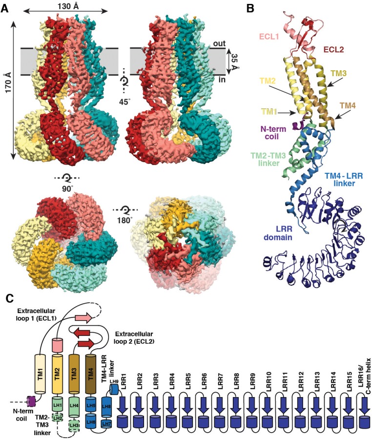
Figure 1.

Figure 1—figure supplement 1. SWELL1 overexpression in cells lacking other LRRC8 subunits produces small DCPIB-sensitive swelling-induced whole cell currents.
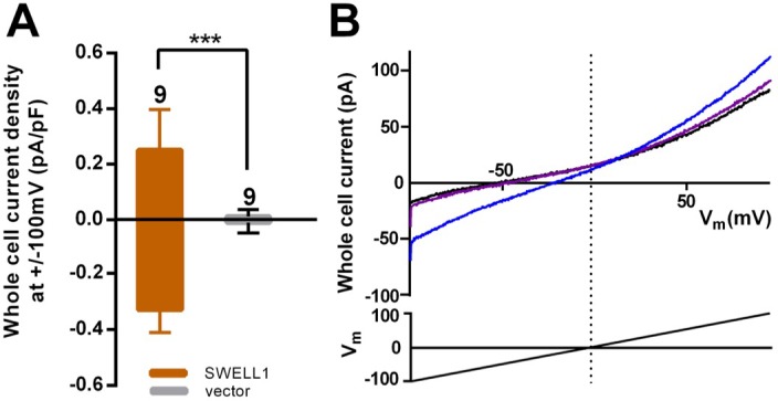
Figure 1—figure supplement 1.

(A) HeLa LRRC8-/- cells were transfected with SWELL1-pIres-GFP or empty pIres-GFP vector and tested for hypotonic (230 mOsm/kg) solution-induced currents using voltage ramp protocols (Qiu et al., 2014). Whole cell currents were measured at −100 and +100 mV before and 5–7 min after hypotonic challenge and normalized to cell capacitance. Shown are means ± s.e.m. for the number of cells indicated from 3 separate experiments. (B) Hypotonic solution-induced currents (blue) were blocked by DCPIB (20 μM) (purple); ramp-induced current in isotonic solution prior to challenge with hypotonic solution is shown in black. Traces were filtered at 1900 Hz and averaged (ten sweeps each). Extracellular solution (in mM): 90 NaCl, 2 KCl, 1 MgCl2, 1 CaCl2, 10 HEPES, 110 mannitol (isotonic, 300 mOsm/kg) or 30 mannitol (hypotonic, 230 mOsm/kg), pH 7.4 with NaOH. Recording pipettes were filled with intracellular solution containing (in mM): 133 CsCl, 5 EGTA, 2 CaCl2, 1 MgCl2, 10 HEPES, 4 Mg-ATP, 0.5 Na-GTP (pH 7.3 with CsOH; 106 nM free Ca2+) and had resistances of 2–3 MΩ.

Figure 1—figure supplement 2. Purification of SWELL1-FLAG.
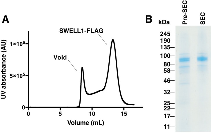
Figure 1—figure supplement 2.

(A) Gel filtration trace of SWELL1-FLAG after affinity purification. (B) SDS-PAGE gel of SWELL1-FLAG. Lane 1 is eluate after affinity purification (Pre-SEC) and lane 2 is combined fractions from SWELL1-FLAG peak post size exclusion. Expected molecular weight is 94 kDa.

Figure 1—figure supplement 3. Cryo-EM data collection.
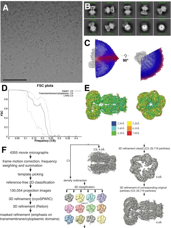
Figure 1—figure supplement 3.

(A) Representative aligned micrograph (scale bar, 100 µm). 4355 movies were collected of SWELL1-FLAG in vitreous ice. (B) Representative 2D classes showing range of orientations of SWELL1-FLAG particles. (C) Euler distribution for final map. Applied C3 symmetry was used, thus only one third of the sphere is shown. (D) Fourier shell correlation between two independently refined cryo-data half sets. (E) Local resolution estimates of the final reconstruction. (F) Data-processing flow chart.

Figure 1—figure supplement 4. Model-to-map fit of electron density.
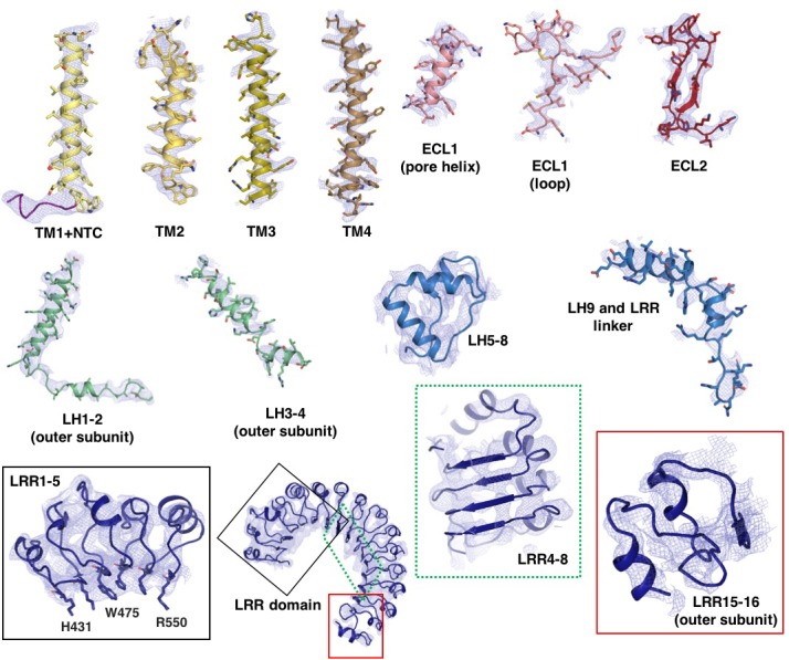
Figure 1—figure supplement 4.

Selected regions of the model are shown with superimposed electron density. Density is derived from the final C3 symmetry-imposed map sharpened with a b-factor of −110 Å2. The cytosolic loop helices (LH1-4) and LRR 15–16 are from the outer subunit.

Figure 1—figure supplement 5. SWELL1 subunit pair and structural homology of SWELL1 structure to connexin-26 and innexin-6 structures.
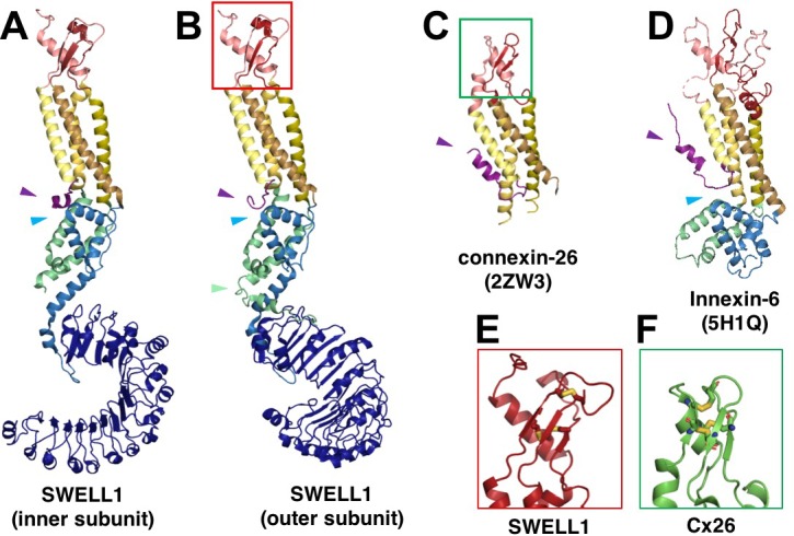
Figure 1—figure supplement 5.

(A–B) Comparison of the SWELL1 ‘inner subunit’ and ‘outer subunit’ in a dimer pair. In the inner subunit, the TM2-TM3 linker contributes two long parallel helices (LH1 and LH4) with a flexible loop of res175-231 between them, while the outer subunit has an additional pair of parallel helices (LH2 and LH3) kinked to place them between the two subunits, though still with an unresolved loop of residues 176–213 between them. LH2, corresponding to residues R214-E236, sits atop of the LRR in the outer subunit (green arrow). Additionally, there is a rotation of the LRR domains relative to the TM domains that allows them to dimerize. (A–D) The LRRC8 family has been shown to be related by weak sequence homology to pannexins (Abascal and Zardoya, 2012), which are in turn related by structural homology to connexins and innexins. The transmembrane helices (TM1-4) of SWELL1 share the same order and arrangement as those of connexin-26 (Cx26; PDB: 2ZW3) (Maeda et al., 2009) and innexin-6 (PDB: 5H1Q) (Oshima et al., 2016) with TM1 closest to the central axis and TM3 and TM4 facing the membrane environment. An N-terminal helix in both Cx26 and innexin-6 creates a pore funnel that forms the narrowest constriction of the channel (Maeda et al., 2009; Oshima et al., 2016), which may be recapitulated in SWELL1 (purple arrows) (E–F) The extracellular loops of SWELL1 (red box) share 3 structurally conserved disulfide bonds with connexin-26 (green box), as well as a three-strand antiparallel beta sheet composed of the antiparallel beta hairpin of ECL2, and a beta strand from ECL1 (Maeda et al., 2009). (A–B, D) In SWELL1, cytoplasmic core formed by the intracellular linker domains share the same topology with portions of the C-terminal domains of distantly related innexin-6 (blue arrows) (Oshima et al., 2016).

Figure 1—figure supplement 6. Alignment of LRRC8 subunits (res 1–435).
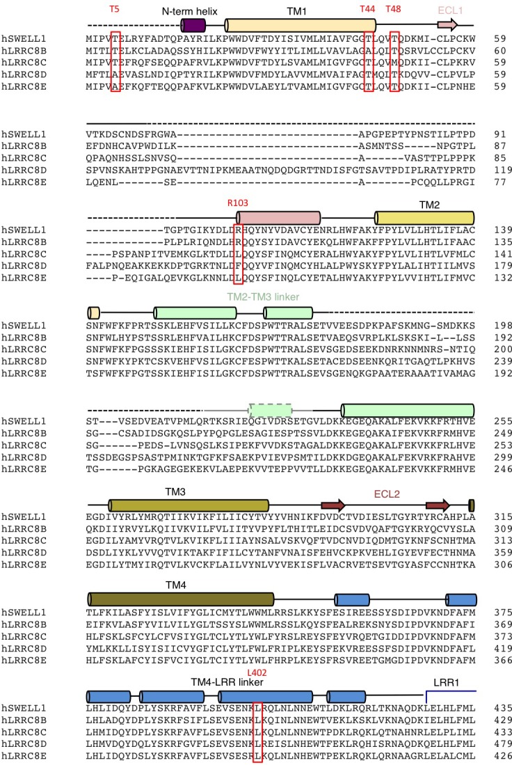
Figure 1—figure supplement 6.

Human LRRC8 family aligned using ClustalW. Secondary structure and domain assignments are annotated above. Residues mentioned in text are boxed red.

Figure 1—figure supplement 7. Alignment of LRRC8 subunits (res 436–810).
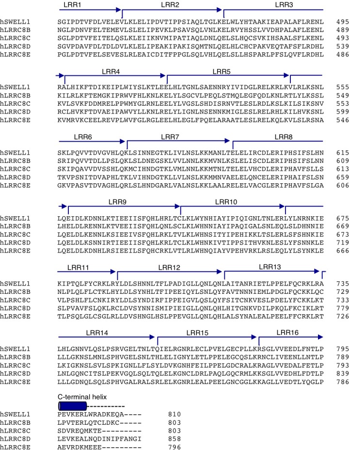
Figure 1—figure supplement 7.

Human LRRC8 family aligned using ClustalW. Secondary structure and domain assignments are annotated above. Residues mentioned in text are boxed red.