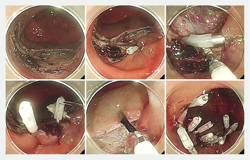
Fig. 1.

Mucosa-submucosa clip closure method. (Upper left) A mucosal defect after colonic endoscopic submucosal dissection. (Upper middle) The first endoclip was placed at the edge of the mucosal defect. Each arm of the endoclip hooked mucosa and submucosa, respectively. The direction of the endoclips was parallel to the short axis of the defect. (Upper right) The second endoclip was also applied in this way. (Lower left) The third endoclip was placed and the mucosal defect was significantly reduced in size. (Lower middle) The fourth clip hooked both sides of the mucosa. (Lower right) Additional endoclips were placed to achieve complete closure.
