
Figure 7. Serotonin is required for the cell-non-autonomous UPRmt induction upon neuronal Wnt/EGL-20 expression.
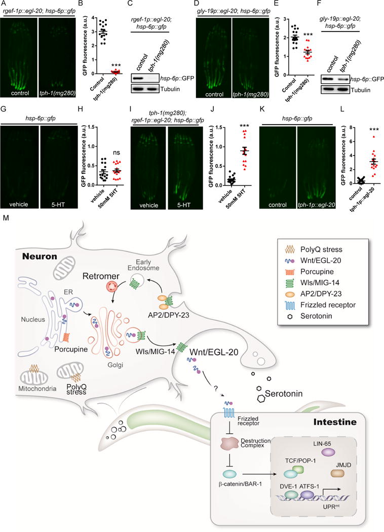
(A) Representative photomicrographs of rgef-1p∷egl-20; hsp-6p∷gfp animals in WT or tph-1 background.
(B) Quantification of hsp-6p∷gfp expression. The genotypes are as in (A).
(C) Immunoblot of hsp-6p∷gfp expression. The genotypes are as in (A).
(D) Representative photomicrographs of gly-19p∷egl-20; hsp-6p∷gfp transgenic animals in WT or tph-1 background.
(E) Quantification of hsp-6p∷gfp expression. The genotypes are as in (D).
(F) Immunoblot of hsp-6p∷gfp expression. The genotypes are as in (D).
(G) Representative photomicrographs of hsp-6p∷gfp expression in animals treated with vehicle control or 50mM serotonin (5-HT).
(H) Quantification of hsp-6p∷gfp expression. The genotypes are as in (G).
(I) Representative photomicrographs of hsp-6p∷gfp expression in neuronal Q40; tph-1 expressing animals treated with vehicle control or 50mM 5-HT.
(J) Quantification of hsp-6p∷gfp expression. The genotypes are as in (I).
(K) Representative photomicrographs of hsp-6p∷gfp reporter animals in WT and tph-1p∷egl-20 background.
(L) Quantification of hsp-6p∷gfp expression. The genotypes are as in (K).
(M) Model of the mitokine signaling pathway. Q40 specifically binds to mitochondria in neurons, initiating a signaling cascade across tissues that requires retromer-dependent Wnt secretion, canonical Wnt signaling, serotonin, and functional components of the UPRmt to ensure cell-non-autonomous UPRmt induction in peripheral tissues.
*** p< 0.0001 via t-test. Error bars, SEM. n ≥ 15 worms.
See also Figure S7.