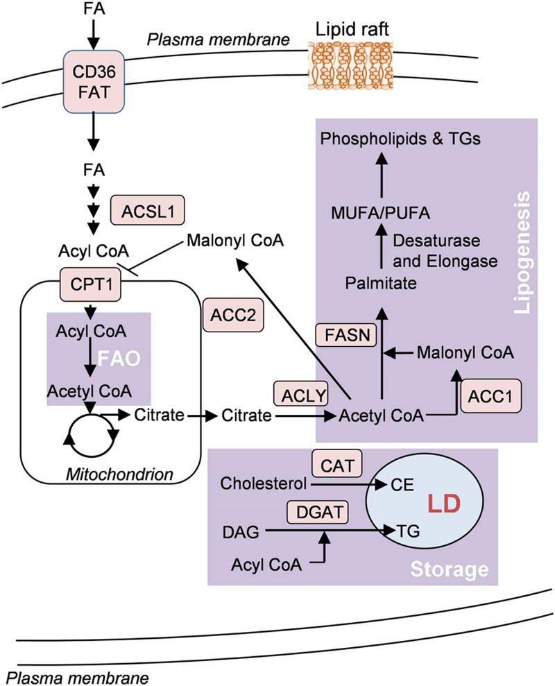
Figure 1.
A simplified schematic representation of lipid metabolism in tumor cells. Tumor cells show altered lipid metabolic networks involving catabolism (fatty acid oxidation (FAO)), biosynthesis pathways (de novo lipogenesis) and storage as lipid droplets (LDs). Cancer cells show increased fatty acid (FA) uptake by fatty acid translocase (FAT), CD36, which then undergo β-oxidation in the mitochondrial matrix. Acetyl CoA, the end product of FAO pathway, can then either enter TCA cycle or may be transported to the cytosol in the form of citrate for fatty acid synthesis. Excess acyl CoA can undergo esterification with cholesterol or diacylglycerol (DAG) to form cholesterol ester (CE) or triacylglycerol (TG) and is stored in the form of lipid droplets.

