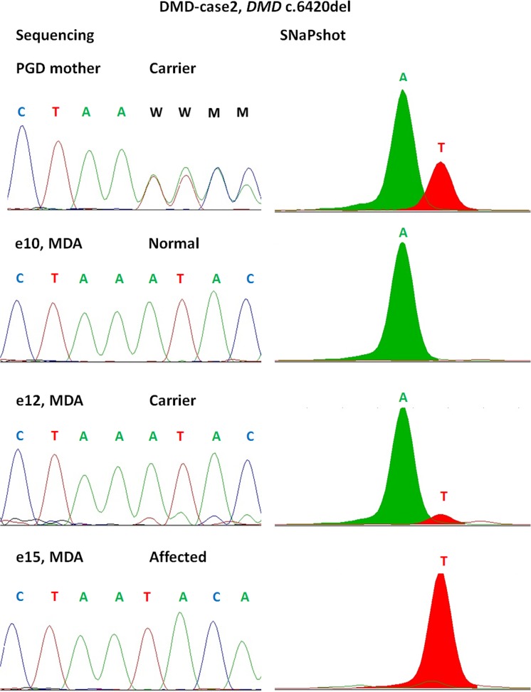
Fig. 3.
Comparison of STR sizing (A) and SNaPshot (B). Results shown for DMD-case 2 variant locus (DMD c.6420del). Whole genome amplification performed by multiple displacement amplification (MDA) technique for three embryos shown (e10, e12, e15). Profiles completely match between two technologies, and partial allelic dropout is visible on both profiles for the heterozygous embryo (e12). PGD mother (first upper panel) shows locus control performed on gDNA

