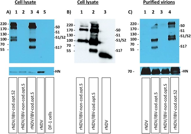
Figure 2.
Western blot analysis of rNDVs expressing S or S2 protein of IBV. The expression of codon optimized S, and S2 proteins and non-codon optimized S protein of IBV were detected by Western blot analysis in infected DF-1 cell lysates, using a chicken polyclonal anti IBV serum (A-upper panel & B). The panel B was added to show the expression of non-codon optimized S protein clearly, since the expression of non-codon optimized S protein was not clear in panel A. For the codon optimized S protein of IBV expressed from rNDV (A-lane 3 and B-lane 2) two bands on top (~170–220 kDa) represent uncleaved S protein (S0) or polymeric forms of S1 or S2 protein. The ~130 kDa band, ~95 kDa band and ~60 kDa band represent S2 or S1 subunit of cleaved S protein. In the case of non-codon optimized S protein expressed from rNDV (2A-lane 2 and B-lane 1), two bands on top (~170–220 kDa) represent uncleaved S protein (S0) or polymeric forms of S2 or S1 protein and ~95 kDa band represents S2 or S1 subunit of cleaved S protein. In the case of rNDV/IBV- S2 (A-lane 1), there are two bands (~170–220 kDa) on top, representing polymeric forms of S2 protein, the ~105 kDa and the ~95 kDa band representing S2 subunit. Lane 4 of panel A and lane 3 of panel B represent rNDV as control. Lane 5 of panel A represents non-infected DF-1 cells. The incorporation of codon optimized S2 and S proteins and non-codon optimized S protein of IBV in NDV particles were detected by Western blot (C-upper panel). Two bands (~170–220 kDa) on top represent uncleaved S protein (S0) or polymeric forms of S2 or S1 protein (C-lane 2). The ~95 kDa and the ~60 kDa band represent S2 or S1 subunit of cleaved S protein (C-lane 2). The two bands (~170–220 kDa) on top represent polymeric forms of S2 protein, the ~105 kDa band and the ~95 kDa band represent S2 subunit(C-lane 4). The lane 1 and 3 of panel C represent purified rNDV control and purified rNDV expressing non-codon optimized S protein, respectively. A monoclonal anti-NDV/HN antibody was used to detect the 70 kDa of HN protein of NDV in DF-1 cell lysates (A-lower panel); rNDV/S2 (lane 1), rNDV/IBV-non-cod.opt.S (lane 2), rNDV/IBV- cod.opt.S (lane 3), rNDV (lane 4) and incorporated in NDV particles (C-lower panel); rNDV (lane 1), rNDV/IBV-cod.opt.S (lane 2), rNDV/IBV-non-cod.opt.S (lane 3) and rNDV/S2 (lane 4). The full-length gels are presented in supplementary Figure S1.

