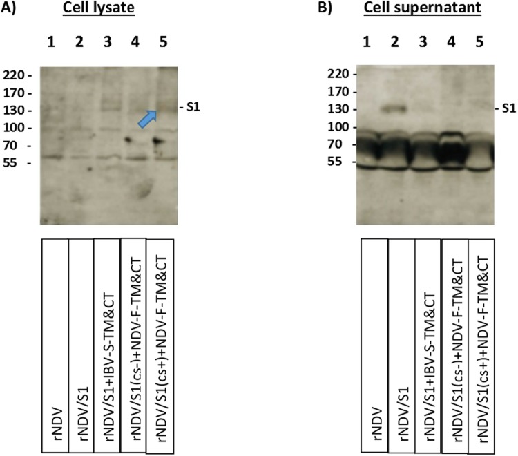
Figure 3.
Western blot analysis of rNDV expressing S1 protein of IBV. The expression of codon optimized S1 protein of IBV expressed from four individual rNDVs expressing four different expression cassettes of S1 protein were detected using Western blot in cell lysates (A) and cell supernatant (B) of infected DF-1 cells infected with rNDVs, using a chicken polyclonal anti IBV serum. The lanes 1–5 represent cell lysates of rNDV, rNDV/S1, rNDV/S1 + IBV-S-TM&CT, rNDV/S1(cs−)+NDV-F-TM&CT, rNDV/S1(cs+) + NDV-F-TM&CT, respectively. A ~130 kD band represent expression of S1 protein by rNDV/S1 + IBV-S-TM&CT, rNDV/S1(cs−) + NDV-F-TM&CT and rNDV/S1(cs+) + NDV-F-TM&CT in infected DF-1 cell lysate (A lanes 3–5) and rNDV/S1 in infected DF-1 cell supernatant (B-lane 2). The full-length gel is presented in Supplementary Figure S1.

