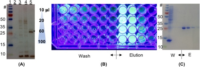
Figure 3.
Expression, protein trans splicing, and purification of eGFP- NpuDnaEΔC16 in E. coli prior to light triggered release. (A) WB using anti-polyhistidine antibody for the detection in the soluble fraction of E. coli lysate. Lanes: (1) molecular weight markers; (2) eGFP- NpuDnaEΔC16 (not induced: negative control); (3) eGFP- NpuDnaEΔC16 + peptide 1; (4) eGFP- NpuDnaEΔC16 + peptide 3 + 1 mM ZnCl2; (5) eGFP- NpuDnaEΔC16 + peptide 3 + 1 mM TCEP. (*) indicate the mass of eGFP-His6. (B) Elution of the soluble protein fractions after Ni-NTA affinity chromatography, using UV-light for visualization, of the products of the protein trans splicing of eGFP- NpuDnaEΔC16 peptide 1. (C) SDS-PAGE stained with CBB of the first four washing steps and the elution steps. The uncropped images can be seen in Fig. S7.

