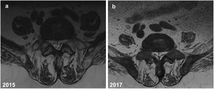
Fig. 1.
a, b A 75-year-old man with advanced metastatic prostate cancer. Axial T2-weighted MR images of the lower lumbar spine a 2015 (1.5 Tesla, TR 4671 ms, TE 92 ms, slice thickness 4 mm) and b 2017 (1.5 Tesla, TR 2260 ms, TE 110.76 ms, slice thickness 4 mm). By comparison, images show increasing abundant epidural fat compressing the thecal sac with obvious tapering, especially b 2017 image demonstrating a “Y” sign indicative of high-grade thecal sac compression

