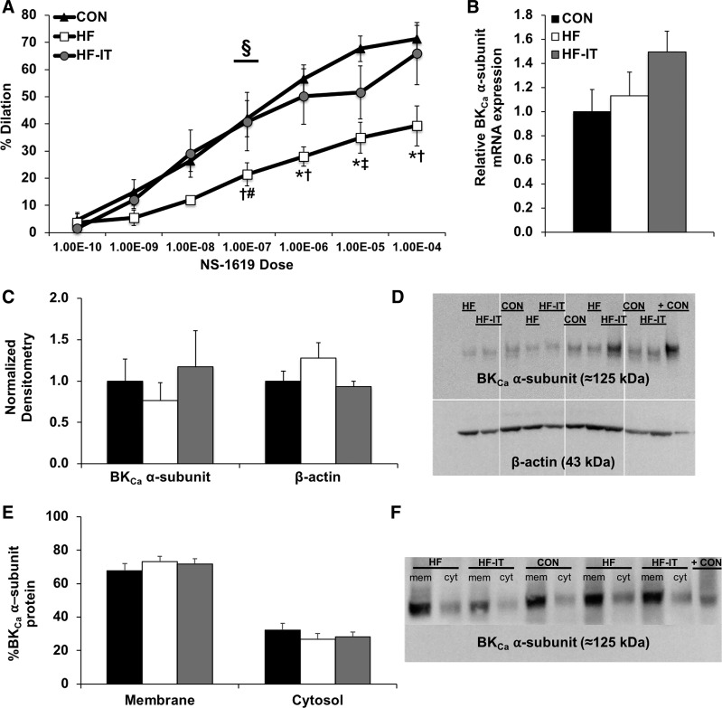
Fig. 4.
In vitro coronary arteriole functional responses to large-conductance Ca2+-activated K+ (BKCa) channel α-subunit activation in nonsham sedentary control (CON), aortic-banded heart failure sedentary (HF), and aortic-banded heart failure interval exercise-trained (HF-IT) animals. A: exercise training prevents the decreased vasodilation in isolated coronary arterioles observed in HF animals after activation of the BKCa channel α-subunit by NS-1619 (repeated-measures ANOVA, P < 0.05). B and C: biochemical analysis of isolated coronary arterioles show no differences in BKCa channel α-subunit mRNA levels (B) or protein (C). D: representative Western blots of BKCa channel α-subunit and β-actin (loading control) arteriole protein levels. + CON, positive control – rat brain. A separate group of animals not included in the analysis of the present report were run on the original gel. Since these samples are not relevant to the present study, they have been removed from the gel as indicated by white dividing lines. E: biotinylation analysis reveals no differences in the cellular distribution of the BKCa channel α-subunit in denuded right coronary artery samples. F: representative Western blot of membrane (mem) and cytosolic (cyt) BKCa channel α-subunit protein levels in the right coronary artery. + CON, positive control – CON arteriole sample from D. §Interaction effect: Group × Dose (P < 0.05); *post hoc vs. CON (P < 0.05); †post hoc vs. HF-IT (P < 0.05); #post hoc vs. CON (P < 0.10); ‡post hoc vs. HF-IT (P < 0.10). n = 6, 5, and 6 for CON, HF, and HF-IT, respectively, for A; n = 6, 6, and 7 for CON, HF, and HF-IT, respectively, for B–F.

