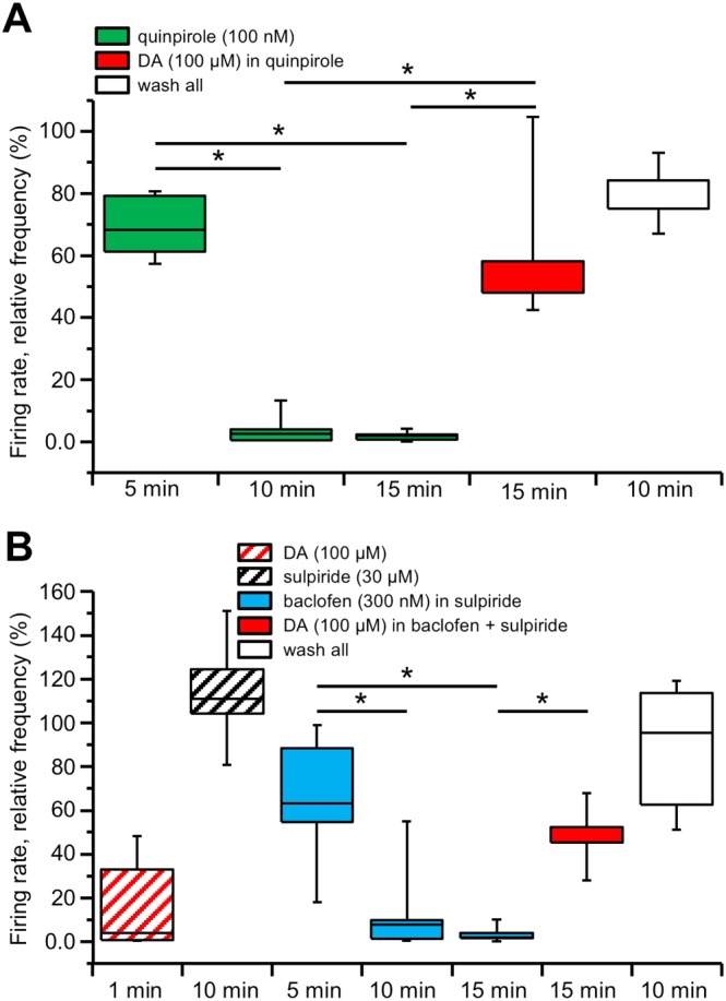
Figure 2.

DIR relies on the presence of dopamine (DA) but not through D2 receptor stimulation. Box‐and‐whisker plots in both panels represent the median values of the firing rate of all the neurons recorded in each slice with MEA, normalized (in %) to control firing rate and measured at the times indicated at the bottom. The centre lines denote medians, edges are upper and lower quartiles and whiskers show minimum and maximum values. (A) Perfusion with the D2 receptor agonist quinpirole (100 nM) induced a pronounced inhibition of the firing rate that reached a plateau within 10 min and remained inhibited at 15 min. Conversely, the firing rate was largely restored in response to a 15 min perfusion with dopamine (100 μM), in the continuous presence of 100 nM quinpirole, such that the relative firing rates at 15 min in quinpirole and at 15 min in quinpirole and dopamine were significantly different (* P < 0.05, one‐way repeated measures ANOVA with Bonferroni's post hoc test, n = 5 slices). Further recovery was attained 10 min after washout of all applied drugs. (B) An initial challenge with a brief 1–2 min perfusion with dopamine (100 μM) inhibited the firing rate of the recorded neurons, confirming their dopaminergic identity. Following this brief dopamine challenge, the slice was perfused with the D2 receptor antagonist sulpiride (30 μM) and kept in the medium thereafter. Perfusion with baclofen (300 nM) induced a pronounced inhibition of the firing rate, reaching a plateau at 15 min. The firing rate was largely restored in response to a 15 min perfusion with dopamine (100 μM), in the continuous presence of 300 nM baclofen, such that the relative firing rates at 15 min in baclofen and at 15 min in baclofen and dopamine were significantly different (* P < 0.05, one‐way repeated measures ANOVA with Bonferroni's post hoc test, n = 5 slices). Further recovery was attained 10 min after washout of all applied drugs.
