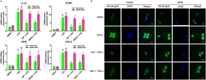
Figure 7.

Knockdown of hPXR attenuated IMP‐mediated suppression of pro‐inflammatory gene expression in LS174T cells. (A) Cells were transiently transfected with hPXR siRNA and control non‐silencing siRNA. After transfection for 48 h, the cells were treated with DMSO, IMP (25 μM) or rifampicin (RIF; 10 μM) for another 48 h, followed by additional exposure to LPS (2000 ng·mL−1) for 24 h. The mRNA expression levels of IL‐1β, ICAM1, iNOS and COX‐2 were determined by qRT‐PCR. Results are expressed as fold changes compared with DMSO control. Data are expressed as mean ± SEM from five independent experiments. # P < 0.05 compared with DMSO group. * P < 0.05 compared with the same treatment group. (B) LS174T cells were transiently transfected with hPXR siRNA and control non‐silencing siRNA for 24 h and then treated with DMSO, IMP (25 μM) or rifampicin (10 μM) for another 48 h, followed by an additional incubation with or without TNF‐α (20 ng·mL−1) for 24 h. NF‐κB p65 localization was performed using immunofluorescence staining and observed under a confocal laser scanning microscope (magnification: 630×) using an anti‐NF‐κB p65 antibody (1:50) followed by an Alexa Fluor 488‐conjugated detection antibody.
