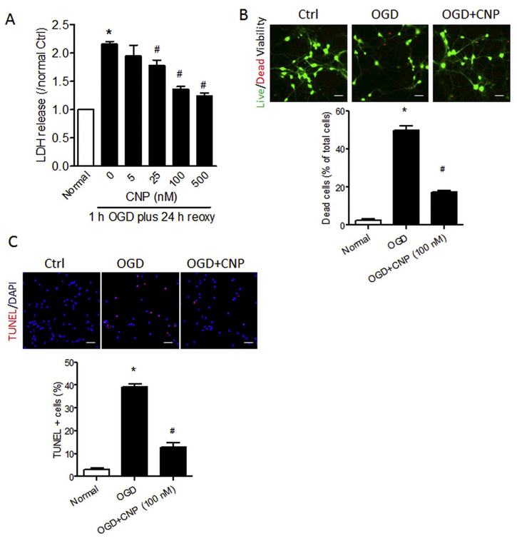
Fig. 3.
Recombinant CNP protects primary cortical neurons from OGD insult. Primary cortical neurons were incubated with 0, 5, 25, 100 or 500 nM of recombinant CNP for 6 h, and then exposed to OGD treatment for 1.0 h followed by reoxygenation for 24 h. LDH release (A), cell viability (B), and TUNEL staining (C) were performed at the end of reoxygenation. (B) Representative images of live (green) and dead (red) cells and quantification of the percentage of dead cells over total cells with recombinant CNP treatment (100 nM) after OGD/reoxygenation. Scale bar, 40 μm. (C) Representative images of TUNEL (red) staining and quantification of the TUNEL-positive cells over total cells with recombinant CNP treatment (100 nM) after OGD/reoxygenation. Data are expressed as mean ± SEM. n = 3 independent experiments. *, p < 0.05 vs. Normal. #, p < 0.05 vs. OGD. ANOVA following by Newman-Keuls post hoc test. (For interpretation of the references to color in this figure legend, the reader is referred to the web version of this article.)

