Figure 6.

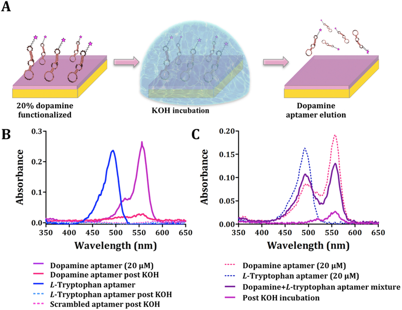
Elution of aptamers following substrate capture. (A) Schematic (not to scale) of incubation of dopamine-functionalized substrates with dopamine aptamers. Unbound sequences were rinsed from substrates. Captured aptamers were eluted by treatment with KOH. (B) Representative UV-vis spectra (N=3) showing the absorbance spectra of eluted aptamers based on AlexaFluor® 546 and AlexaFluor® 488 emission wavelengths for dopamine (correct and scrambled) and L-tryptophan aptamers, respectively. Control experiments with the L-tryptophan aptamer and the scrambled dopamine aptamer showed negligible nucleic acid elution. (C) Representative UV-vis spectra (N=3) for selective elution of dopamine aptamers from mixtures with L-tryptophan aptamers (1:1 ratio).
