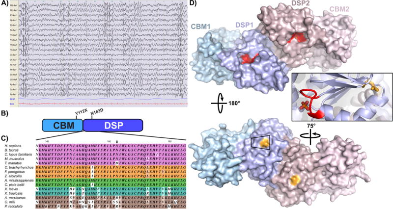
Fig. 1. Phenotypic and genotypic characterization of an unusual case of LD.

A) EEG analysis of the patient when she was 28 years old. The EEG shows a slow background activity with bilateral spike and spike and wave discharges. B) Domain structure of the EPM2A gene encoding laforin. Y112X maps to the end of the CBM, while N163D maps to the DSP domain. C) Representative partial alignment of DSP domain sequences, colored by vertebrate class. 43% of residues are strictly conserved based on our analysis (See supplemental file S2 for full alignment) while N163 is substituted with an aspartate, serine or arginine. D) The position of the mutations present in the LD patient were mapped in the laforin structure described recently (Raththagala et al., 2015) (PDB 4RKK) using PyMol software (DeLano Scientific LLC, USA). The two subunits of the dimer are shown in shades of blue and pink; the CBM is shown as a lighter shade than the DSP domain. The classic DSP catalytic motif (CX5R) is shown in red on the front face of the dimer, and the bound phosphate is partially obscured from view. N163 (shown in light orange) maps to the back of the dimer, opposite to the catalytic cleft (red) and bound phosphate.
