Figure 3.
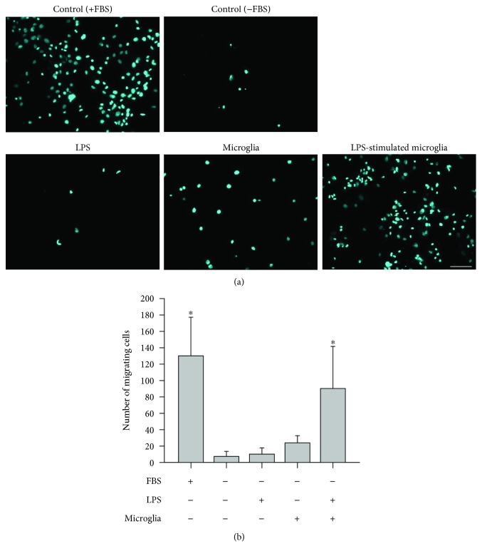
Migration assays with rat bone marrow-derived mesenchymal stem cells (rBM-MSCs) cocultured with LPS-stimulated microglia. (a) Images of migrating rBM-MSCs. Migration of rBM-MSCs was investigated in 5 different conditions: media containing foetal bovine serum (FBS) (positive control), media without FBS (negative control), LPS-treated, cocultured with microglia, and coculture with LPS-stimulated microglia. Migrating cells are cyan in colour (10x magnification). Representative images of migration for each condition are shown. Scale bar: 100 μm. (b) Quantification of migrating cells was performed by counting coloured dots in the images. Data represent the mean of ten random 372.23 mm2 (710.52 μm × 532.38 μm) microscopic fields (mean ± SD). ∗ p < 0.05 versus negative control (media without FBS).

