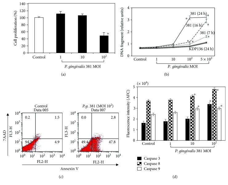
Figure 1.
P. gingivalis infection inhibits cellular proliferation and induces apoptosis in HUVEC. (a) HUVEC were treated with P. gingivalis 381 at an MOI of 1 : 1–1 : 102 for 21 h. Cell viability was determined using a Cell Counting Kit-8 (CCK-8). The data are expressed as the mean ± SEM of 3 different experiments. ∗p < 0.05 versus bacteria-free control cells. (b) HUVEC were treated with P. gingivalis 381 at an MOI of 1 : 1–1 : 5 × 10² for 7, 16, and 24 h, and with KDP136 at an MOI of 1 : 1–1 : 5 × 10² for 24 h. Cellular apoptosis was quantified by DNA fragmentation using the Cell Death Detection ELISAPLUS Kit as described in Materials and Methods. Data are expressed as the mean ± SEM of 3 different experiments. ∗p < 0.05 versus bacteria-free control cells. (c) HUVEC were stained with annexin V-EnzoGold and 7-AAD after treatment with P. gingivalis 381 at an MOI of 1 : 102 for 21 h, and then they were analyzed by flow cytometry. The figure is representative of three experiments with similar results. (d) HUVEC were treated with P. gingivalis 381 at an MOI of 1 : 1–1 : 102 for 16 h. Cell extracts were prepared, and caspase activities were measured. Data are expressed as the mean ± SEM of 3 different experiments. ∗p < 0.05 versus bacteria-free control cells.

