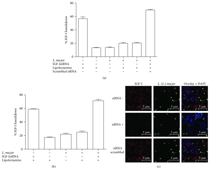
Figure 3.
Expression of Igf-I mRNA upon IGF-I silencing with an siRNA. The percentage decrease in the amount of Igf-I mRNA in RAW 264.7 cells (a) or BALB/c peritoneal macrophages (b) infected with L. major promastigotes that were transfected with 150 μM siRNA, scrambled siRNA, or Lipofectamine alone 6 h after infection is shown. One representative experiment from three independent assays is shown. (c) The detection of IGF-I expression in L. major promastigote-infected RAW 264.7 cells transfected with (siRNA+) or without the IGF-I siRNA (siRNA−) or with a scrambled siRNA using confocal microscopy of immunostaining with a 1 : 75 dilution of an anti-IGF-I antibody (using an Alexa Fluor 546-conjugated secondary antibody; red) and a 1 : 200 dilution of an anti-Leishmania antibody (using an Alexa Fluor 488-conjugated secondary antibody; green) is shown. Nuclei were stained with DAPI (blue). Images were captured using a confocal Leica LSM510 microscope with a 63x oil immersion objective.

