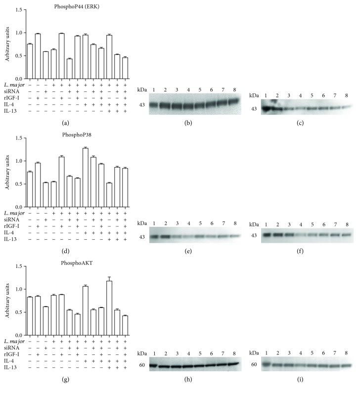
Figure 7.
The effects of siRNA and IL-4 on components of the IGF-I signaling pathways: levels of phosphorylated p44 (ERK), p38 (MAPK), and AKT proteins. Promastigote-infected or noninfected cells transfected with or without IGF-I siRNA were stimulated for 30 minutes with IL-4 (2 ng/mL) and IL-13 (5 ng/mL). Cells were lysed, proteins were separated by 10% SDS-PAGE, and subsequently, Western blotting was performed using anti-phospho-p44 (a, b, and c), anti-phospho-p38 (d, e, and f), and anti-phospho-AKT (g, h, and i) antibodies. Protein bands corresponding to protein expression levels were subject to a densitometric analysis, and the data are expressed in arbitrary units (a, d, and g). A representative blot is shown. (b, e, h) The lanes represented the following: 1: control; 2: RAW; 3: RAW + rIGF; 4: RAW + siRNA; 5: RAW + Lm; 6: RAW + Lm + rIGF; 7: RAW + Lm + IL-4; and 8: RAW + Lm + IL-4 + IL-13. (c, f, i): 1: control; 2: RAW; 3: RAW + Lm + siRNA; 4: RAW + Lm + siRNA + rIGF; 5: RAW + Lm + siRNA + IL-4; 6: RAW + Lm + siRNA + IL-4 + rIGF; 7: RAW + Lm + siRNA + IL-4 + IL-13; and 8: RAW + Lm + siRNA + IL-4 + IL-13 + rIGF. See Materials and Methods for additional details.

