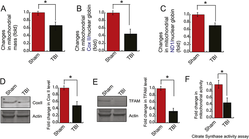
Figure 1: TBI causes a decrease in mitochondrial mass.

(A) Fold changes in mitochondrial mass obtained from staining from MitoTracker Green was monitored in both sham and TBI samples by a spectrofluorometer. It was shown that TBI causes a decrease in mitochondrial mass (n=3–5; one-way ANOVA, p<0.005). (B-C) Mitochondrial DNA quantification by Real-time PCR was represented by mitochondrial COXII (B) and mitochondrial ND1 (C) normalized to nuclear ß-globin isolated from nuclear DNA. It was shown that COXII and ND1 was decreased significantly after TBI. (n=3–5; one-way ANOVA, p<0.005) (D-E) Western blot and quantitative analysis of the expression of TFAM (D) and COXII (E) level in both sham and TBI samples. The band intensity of TFAM (D) and COXII (E) was monitored by Image J. The expression level of TFAM and COXII was decreased significantly after TBI. (n=3–5; one-way ANOVA, *p<0.005).
