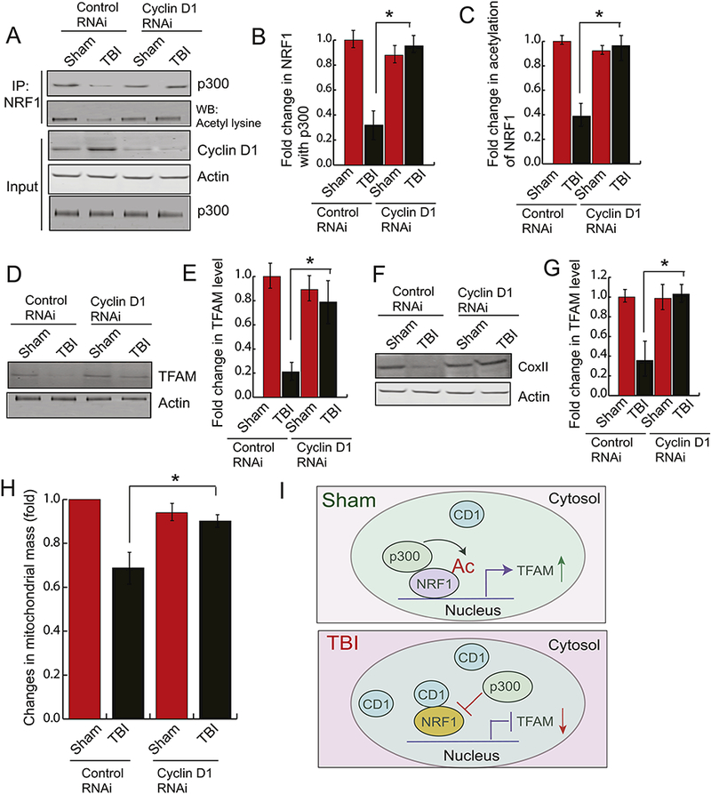
Figure 4: Depletion of Cyclin D1 rescue the loss of mitochondrial mass following TBI.

Cyclin Dl-siRNA was administered into the mouse brain by intranasal delivery before TBI. (A-C) The pericontusional cortex was used to perform immunoprecipitation assay to detect acetylation of NRFl and the interaction between NRF1 and p300. The expression level of p300, cyclin D1 and Actin was monitored by western blot hybridization. The band intensity of the interaction between p300-NRF1 (B) and the level of acetylated NRF1 (C) was monitored by Image J. (n=3–5; one-way ANOVA, *p<0.005). The data shows that depletion of CD1 rescues acetylation of NRF1 and the interaction between p300 and NRF1. (D-E) The pericontusional cortex was used to perform western blot analysis of TFAM (D) and the band intensity of TFAM was monitored by Image J. (n=3–5; one-way ANOVA, *p<0.005) (E) in both sham and TBI mice administered with either control RNAi or CD1 RNAi. The data shows that depletion of CD1 rescues TFAM level after TBI. (F-G) The pericontusional cortex was used to perform western blot analysis of COXI (F), and the band intensity of COXII was monitored by Image J. (n=3–5; one-way ANOVA, *p<0.005) (G) in both sham and TBI mice administered with either control RNAi or CD1 RNAi. The data shows that depletion of CD1 rescues COXII level after TBI. (I) Fold change in mitochondrial mass was monitored in both sham and TBI mice administered with either control RNAi or CD1 RNAi (n=3–5; one-way ANOVA, *p<0.005). The data shows that depletion of CD1 rescues mitochondrial mass after TBI.
