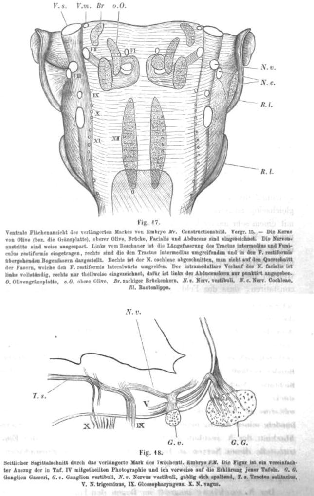
Fig 3.
(Showing His, 1890; Fig. 17) Ventral view of the rhombencephalon of embryo Mr, image of a reconstruction, Ma 15 – The olivary nuclei (or pons), superior olive, pons, facial, abducens are shown). Nerve roots are shown in white. The left shows the intermediate tract and the restiform body, the right shows the arcuate fibers surrounding the intermediate and pass to the restiform body. The right cochlear nucleus is removed to show the fibers that surround laterally the restiform body. The intramedullary course of the facial nerve is completely shown on the left, but only partially on the right, but the abducens is only outlined by a dotted line. G, pons, oO superior olive, Br, dentate potine nucleus; Nv, vestibular nerve; Nc, cochlear nerve; Rl, rhombic lip.

