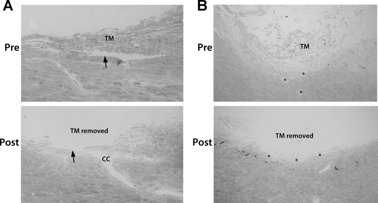
Fig. 1.
Histology showing conventional outflow pathway pre- and post-trabeculotomy in human and porcine eyes. Radial sections (1 µm) from perfusion-fixed human (A) and porcine (B) eyes were taken with the trabecular meshwork (TM) intact and after trabeculotomy. Following trabeculotomy in human eyes, all of the TM and inner wall of Schlemm’s canal were removed, leaving only the outer wall (arrows) and the distal regions present. The images shown were taken from anterior segments from different donors. For porcine eyes, the angular aqueous plexus (AAP) can be seen in both sections (asterisks); however, parts of the AAP are open to the anterior chamber in the post-trabeculotomy section, whereas others are not. CC, collector channel.

