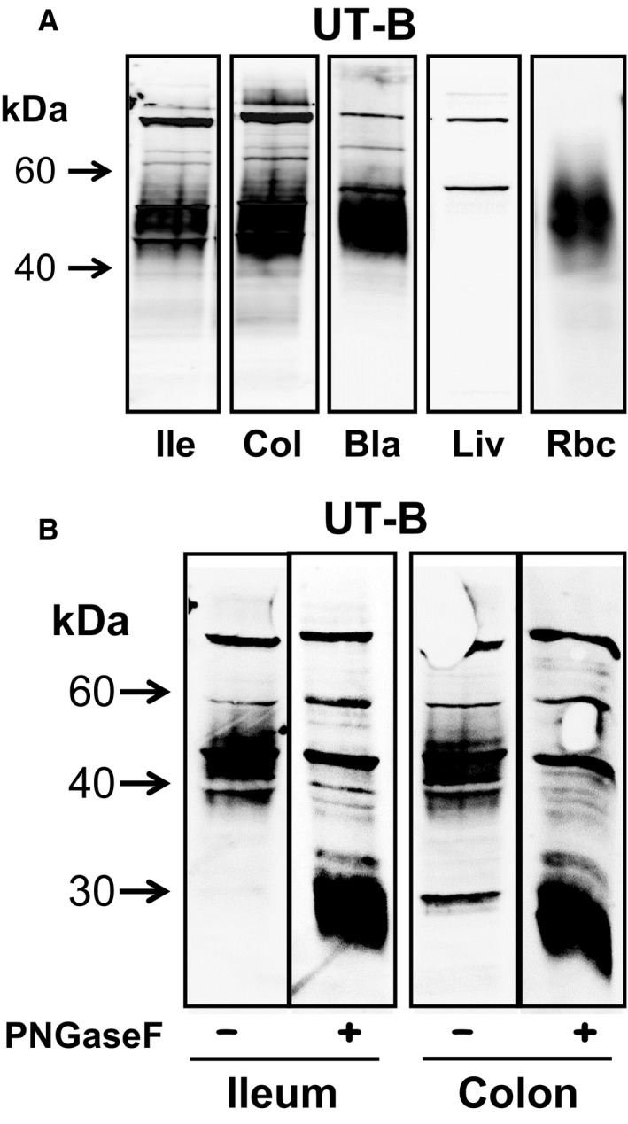
Figure 3.

Western blotting experiments investigating UT‐B protein abundance in various tissues. (A) Using membrane‐enriched protein samples, hUTBc19 antibodies detected strong, smeared 40–60 kDa UT‐B signals in ileum, colon, bladder, and red blood cells. In contrast, no such signal was detected in liver, where only weak, tight bands at ~50 and ~100 kDa were detected. (B) The effect of PNGaseF treatment was investigated on the UT‐B signals in ileum and colon. In both tissues, PNGaseF deglycosylated the 40–60 kDa signal to an unglycosylated core protein at ~30 kDa. Key: Ile = ileum; Col = colon; Bla = bladder, Liv = liver; Rbc = red blood cells; PNGaseF = PNGaseF enzyme; + = PNGaseF treated; − = untreated.
