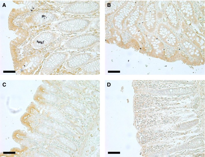
Figure 5.

Immunolocalization showing UT‐B staining of 10 μm transverse sections of different regions of the human colon. (A) Ascending colon (×20 magnification), (B) Tranverse colon (×20 magnification, (C) Descending colon (×20 magnification), and (D) Sigmoid colon (×20 magnification). All scale bars represent 50μm.
