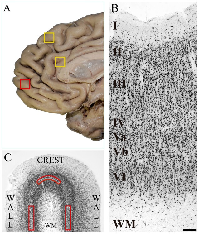
Figure 1.

(A) Medial surface of the human frontal lobe. The yellow boxes show the cortical regions in which von Economo neurons (VENs) have been identified in previous studies: dorsomedial area 9 and anterior cingulate cortex. The red box shows the place where we took tissue samples in the present study, which is located on the medial surface of the frontal pole. (B) Microphotograph of a tissue section of human medial area 10 immunostained with anti-NeuN from the pial surface to the white matter. In the image, the cortical layers and the sublamination of layer V are indicated. The density of pyramidal cells decreases in the deeper part of layer V (Vb), and conversely, the soma area increases. Magnification 10×, scale bar = 200 μm. (C) Diagram of a tissue section stained with anti-NeuN showing the crest and walls of a cortical gyrus. The red boxes show the approximate location of the grids (1500 μm wide × 300 μm thick) for the neuronal counts on sublayer Vb. WM: white matter.
