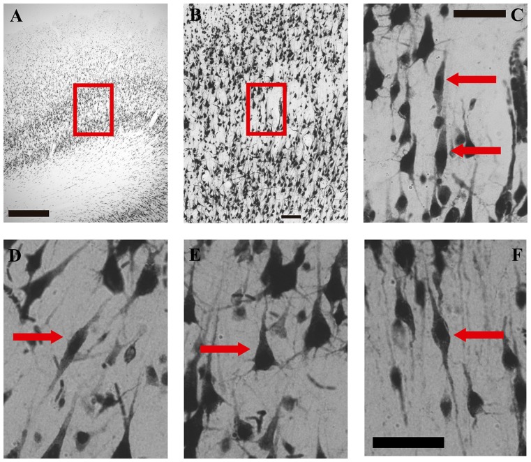
Figure 2.
Microphotographs of VENs, pyramidal and fusiform cells in the human medial frontopolar cortex. (A–C) Correspond to photographs taken at different magnifications of the same sector of one of the walls of a gyrus. (D–F) are high-magnification photographs (40×) of a VENs and a pyramidal cell of sublayer Vb, and of a fusiform cell of layer VI. (A) Portion of a tissue section where VENs were found in the wall of a gyrus. The red box shows the sector located between layers IV and VI which will be enlarged on image (B). Magnification 2.5×, scale bar = 500 μm. (B) Microphotograph illustrating a magnification of the red box of image (A). Magnification 10×, scale bar = 100 μm. (C) Microphotograph illustrating a magnification of the red box in image (B). The red arrows indicate two NeuN-positive cells with the typical morphology of VENs identified in sublayer Vb of the medial frontopolar cortex of humans. Magnification 40×, scale bar = 50 μm. (D) Photomicrograph illustrating a VEN of sublayer Vb indicated by the red arrow. (E) Microphotograph illustrating a pyramidal cell of sublayer Vb indicated by the red arrow. (F) Microphotograph illustrating a fusiform cell of layer VI indicated by the red arrow. Scale bar = 50 μm for images (D–F).

