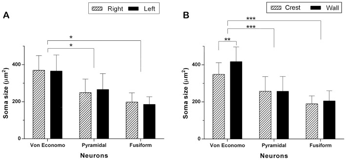
Figure 3.
Comparison of the soma area of VENs, pyramidal and fusiform cells. (A) Neuronal size per hemisphere. There were no statistically significant inter-hemispheric differences in the area of VENs, pyramidal and fusiform cells. The area of the soma of VENs is greater with respect to that of pyramidal and fusiform cells (*p < 0.05). (B) Neuronal area per gyrus portion. There are differences in the area of VENs according to their regional location, being greater in the walls than in the crests of the gyri (**p < 0.01). VENs were larger when compared to pyramidal and fusiform cells, in both portions of the gyri (***p < 0.001). In the latter, there were no differences related to their location along a gyrus.

