
FIGURE 5.
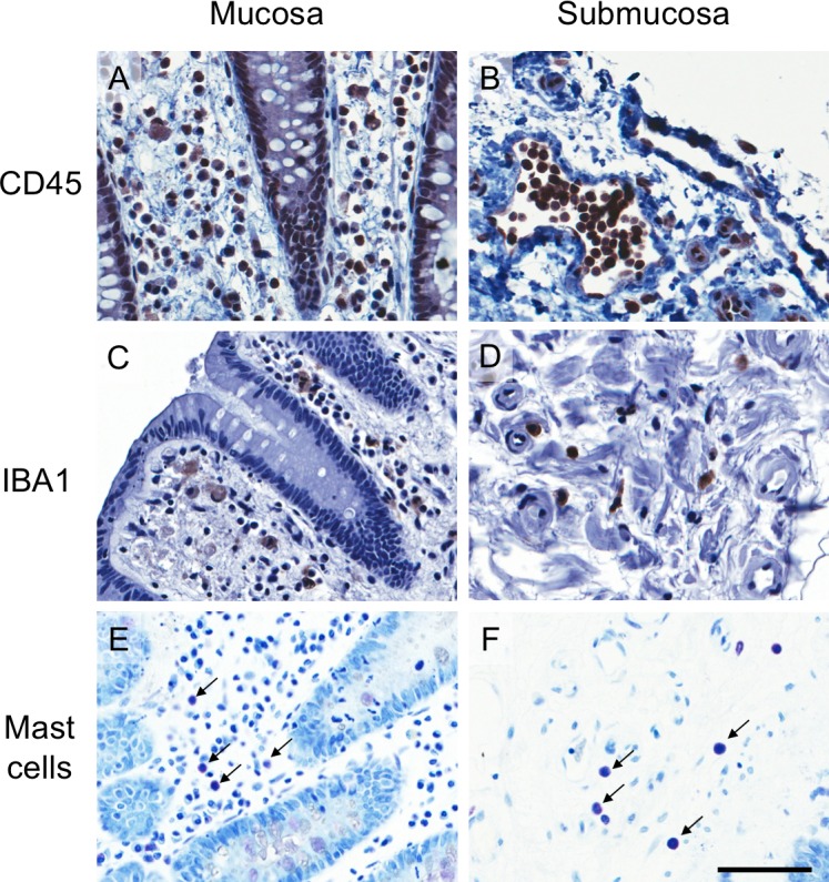
Images of CD45 immunoreactive (IR), IBA1-IR and toluidine blue stained cells from a representative incubation control tissue. (A,B) CD45-IR leukocytes, (C,D) IBA1-IR macrophages and monocytes, and (E,F) toluidine blue stained mast cells in mucosa (left panels) and submucosa (right panels). The scale bar represents 50 μm. All images were taken from the colonic mucosa of a 50-year-old female patient.