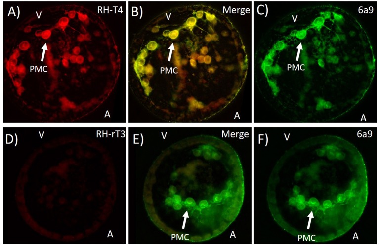
Figure 9.
Colocalization of fluorescently labeled T4 (RH-T4—A), labeled rT3 (RH-rT3—D) with primary mesenchyme cells (PMCs). (B,E) Merged image showing colocalization of fluorescently labeled thyroid hormones and 6a9 antibody for PMCs. Gastrulae were incubated with RH-T4 and RH-rT3 for 30 min prior to fixation in methanol. Following fixation, immunohistochemistry with 6a9 antibody (C,F) was used to stain the membrane of PMCs. RH-T4 binds specifically to the membrane of PMCs, while RH-rT3 does not, suggesting a specific binding site for T4 in the PMCs.

