Fig. 7.
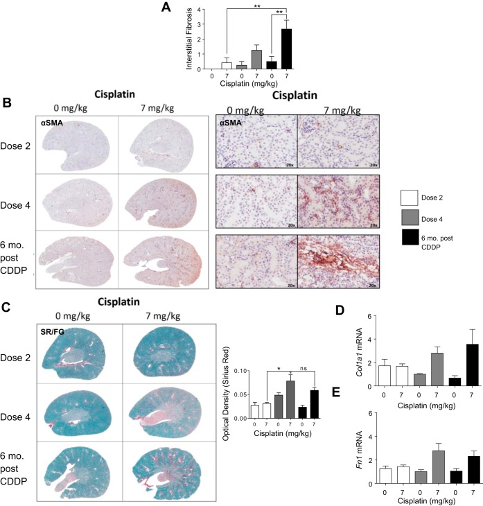
Development of interstitial fibrosis during and after repeated cisplatin (CDDP) treatment. Eight-week-old FVB mice were treated with either saline vehicle or CDDP (7 mg/kg) once per week for 2 wk and euthanized 3 days after dose 2. Another group was treated with either saline vehicle or CDDP (7 mg/kg) once per week for 4 wk and euthanized 3 days after dose 4, or allowed to age for 6 mo following treatment (6 mo post-CDDP). A: interstitial fibrosis levels determined by a renal pathologist. B: presence of myofibroblasts determined by α-smooth muscle actin (α-SMA) IHC. C: levels of total collagen determined by Sirius red/Fast green (SR/FG) staining and optical density quantification of Sirius red staining. Levels of collagen 1 type 1a (Col1a1) (D) and fibronectin (Fn1) (E) measured in kidney cortex via qRT-PCR. Data are expressed as means ± SE; n = 5–10. Statistical significance was determined by 2-way ANOVA followed by Tukey posttest. *P < 0.05, **P < 0.01.

