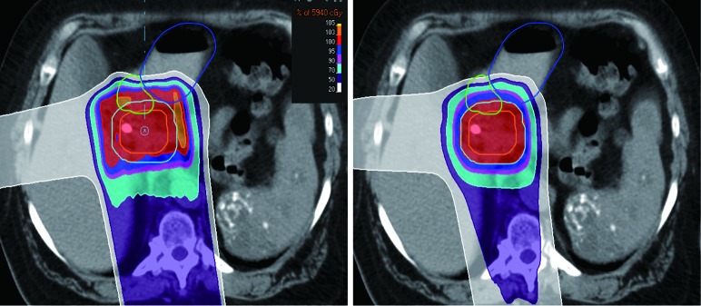
Figure 2.
Dose distribution of passive scattering (PS) plan (left) and pencil beam scanning (PBS) plan (right). Note the tighter high-dose conformality in the PBS plan. IGTV in orange, ICTV in cyan, duodenum in green and stomach in blue. IGTV, internal gross tumor volume; ICTV, internal clinical target volume.

