Fig. 1.
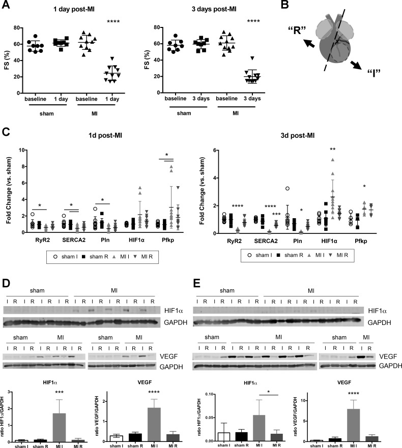
Early response to myocardial infarction (MI) injury included upregulation of hypoxic signaling and decreased cardiac contractility. A: fractional shortening (FS) in sham and MI mice at baseline, 1 and 3 days after MI. B: diagram of tissue dissection after MI. At designated time point, hearts were collected and atria were removed (light gray). Ventricular tissue (dark gray) was then separated into ischemic free wall (I, infarct + border regions of left ventricle) and remainder (R, includes some left ventricle, septum, and right ventricle). Ischemic tissue was identified by tissue blanching, and the surrounding border region was included in this fraction. Sham animals were dissected in a similar manner with the free wall considered the “ischemic” fraction and remaining tissue as “remainder.” Circled area indicates infarcted region of left ventricle, below the suture ligating the left anterior descending artery. C: semiquantitative PCR data for 1 and 3 days (d) after MI. Data shown with means ± SD. For A and C, n = 8–11 mice for each time point except Pfkp at 3 days after MI (n = 4–5). Western blotting and quantification for HIF1α and VEGF in heart tissue lysate at 1 day (D) and 3 days post-MI (E). GAPDH used for loading control and normalization. All analyses were performed with one-way ANOVA with Tukey’s multiple comparisons or Kruskal-Wallis with Dunn’s multiple comparisons. All significant differences indicate comparison to all other fractions unless otherwise denoted. *P < 0.05, **P < 0.01, ***P < 0.001, ****P < 0.0001.

